The spinal cord is characterized by a pronounced segmental structure, reflecting the segmental structure of the vertebrate body. Two pairs of ventral and dorsal roots depart from each spinal segment. The dorsal roots form the afferent inputs of the spinal cord. They are formed by the central processes of the fibers of the primary afferent neurons, the bodies of which are brought to the periphery and are located in the spinal ganglia. The ventral roots form the efferent outlets of the spinal cord. Axons of a and g-motor neurons, as well as preganglionic neurons of the autonomic nervous system. This distribution of afferent and efferent fibers was established at the beginning of the last century and was called the Bell-Magendie law. After transection of the anterior roots on one side, a complete shutdown of motor reactions is observed; but the sensitivity of this side of the body remains. Transection of the posterior roots turns off the sensitivity, but does not lead to the loss of motor reactions of the muscles.
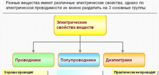
1 - white matter;
2 - gray matter;
3 - back (sensitive) spine;
4 - spinal nerves;
5 - anterior (motor) root;
6 - spinal ganglion
The neurons of the spinal ganglia are simple unipolar, or pseudo-unipolar, neurons. The name "pseudo-unipolar" is explained by the fact that in the embryonic period, the primary afferent neurons originate from bipolar cells, the processes of which then merge. The neurons of the spinal ganglia can be divided into small and large cells. The body of large neurons has a diameter of about 60–120 µm, while in small neurons it ranges from 14 to 30 µm.
Large neurons give rise to thick myelinated fibers. From the small ones, both thin myelinated and unmyelinated fibers begin. After bifurcation, both processes go in opposite directions: the central one enters the dorsal root and, in its composition, enters the spinal cord, the peripheral one enters various somatic and visceral nerves suitable for the receptor formations of the skin, muscles, and internal organs.
Sometimes the central processes of primary afferent neurons extend into the ventral root. This occurs during the trifurcation of the axon of the primary afferent neuron, as a result of which its processes are projected into the spinal cord and through the dorsal and ventral roots.
Of the entire population of dorsal ganglion cells, approximately 60–70% are small neurons. This corresponds to the fact that the number of unmyelinated fibers in the dorsal root exceeds the number of myelinated fibers.
The bodies of neurons in the spinal ganglia do not have dendritic processes and do not receive synoptic inputs. Their excitation occurs as a result of the arrival of an action potential along the peripheral process in contact with the receptors.
Spinal ganglion cells contain high concentrations of glutamic acid, one of the putative mediators. Their surface membrane contains receptors specifically sensitive to g-aminobutyric acid, which coincides with the high sensitivity to g-aminobutyric acid of the central endings of the primary afferent fibers. Small ganglion neurons contain substance P or somatostatin. Both of these polypeptides are also likely mediators released by the endings of primary afferent fibers.
Each pair of roots corresponds to one of the vertebrae and leaves the spinal canal through the opening between them. Therefore, segments of the spinal cord are usually designated by the vertebra near which the corresponding roots emerge from the spinal cord. The spinal cord is also usually divided into several sections: cervical, thoracic, lumbar and sacral, each of which contains several segments. In connection with the development of the limbs, the neural apparatus of those segments of the spinal cord that innervate them has received the greatest development. This was reflected in the formation of cervical and lumbar thickenings. In the area of thickening of the spinal cord, the roots contain the largest number of fibers and have the greatest thickness.
On a transverse section of the spinal cord, the centrally located gray matter, formed by an accumulation of nerve cells, and the white matter fringing it, formed by nerve fibers, are clearly distinguished. In the gray matter, ventral and dorsal horns are distinguished, between which lies an intermediate zone. In addition, in the thoracic segments, there is also a lateral protrusion of the gray matter - the lateral horns.
All neuronal elements of the spinal cord can be divided into 4 main groups: efferent neurons, intercalary neurons, ascending tract neurons, and intraspinal fibers of sensory afferent neurons. Motor neurons are concentrated in the anterior horns, where they form specific nuclei, all of whose cells send their axons to a specific muscle. Each motor nucleus usually extends over several segments. Therefore, the axons of motor neurons innervating the same muscle leave the spinal cord as part of several ventral roots.
In addition to the motor nuclei located in the ventral horns, there are large accumulations of nerve cells in the intermediate zone of gray matter. This is the main nucleus of the intercalary neurons of the spinal cord. Axons of intercalary neurons spread both within the segment and into the nearest neighboring segments.
A characteristic accumulation of nerve cells also occupies the dorsal part of the dorsal horn. These cells form dense interlacings, and the indicated zone is called Roland's gelatinous substance.
The most accurate and systematized idea of the topography of the nerve cells of the gray matter of the spinal cord is given by dividing it into successive layers, or plates, in each of which mainly neurons of the same type are grouped.
Although the layered gray matter typography was initially identified in the cat spinal cord, it turned out to be quite universal and quite applicable to the spinal cord of both other vertebrates and humans.
According to these data, all gray matter can be divided into 10 plates. The very first dorsal plate contains mainly the so-called marginal neurons. Their axons project rostrally, giving rise to the spinothalamic tract. On the edge neurons, the fibers of the Lissauer tract terminate, which is formed by a mixture of primary afferent fibers and axons of propriospinal neurons.
The second and third plates form a gelatinous substance. Two main types of neurons are localized here: smaller and relatively large neurons. Although the bodies of neurons in the second plate have a small diameter, their dendritic ramifications are very numerous. The axons of the second plate neurons project to the Lissauer tract and the spinal cord's own dorsolateral bundle, but many remain within the gelatinous substance. On the cells of the second and third plates, the fibers of the primary afferent neurons terminate, mainly skin and pain sensitivity.
The fourth plate occupies approximately the center of the dorsal horn. The dendrites of layer IV neurons penetrate the gelatinous substance, and their axons project into the thalamus and lateral cervical nucleus. They receive synaptic inputs from the neurons of the gelatinous substance, and their axons are projected to the thalamus and the lateral cervical nucleus. They receive synaptic inputs from neurons of the gelatinous substance and primary afferent neurons.
In general, the nerve cells of the first-fourth plates capture the entire top of the dorsal horn and form the primary sensory area of the spinal cord. The fibers of most of the dorsal-radicular afferents from exteroreceptors are projected here, including skin and pain sensitivity. In the same zone, nerve cells are localized, giving rise to several ascending tracts.
The fifth and sixth plates contain numerous types of intercalary neurons that receive synaptic inputs from the fibers of the dorsal root and descending tracts, especially the corticospinal and rubrospinal tracts.
In the seventh and eighth plates, propriospinal intercalary neurons are localized, giving rise to long axons reaching the neurons of distant segments. Afferent fibers from proprioreceptors, fibers of the vestibulospinal and reticulospinal tracts, axons of propriospinal neurons end here.
The bodies of a- and g-motor neurons are located in the ninth plate. This area is also reached by presynaptic endings of primary afferent fibers from muscle stretch receptors, endings of fibers of descending tracts, cortico-spinal fibers, and axon terminals of excitatory and inhibitory interneurons.
The tenth plate surrounds the spinal canal and contains, along with neurons, a significant amount of glial cells and commissural fibers.
Cells of neuroglia of the spinal cord cover the surface of neurons for a considerable extent, and the processes of the glial cell are directed, on the one hand, to the bodies of neurons, and on the other hand, they often contact with blood capillaries, being intermediaries between nerve elements and their food sources.
The spinal cord transmits signals along the ascending pathways to the suprasegmental levels of the brain, and along the descending pathways it receives commands for action from there. Ascending pathways transmit impulses from proprioceptors along the fibers of the spinobulbar bundles of Gaulle and Burdach and the spinal cerebellar tracts of Govers and Flexigo, from pain and temperature receptors along the lateral spinothalamic tract, from tactile receptors along the ventral spinothalamic tract and partially through the Gaull and Burdach bundles.
Descending paths pass as part of the corticospinal, or pyramidal, tracts and extracorticospinal, or extrapyramidal.
To control the work of internal organs, motor functions, timely receipt and transmission of sympathetic and reflex impulses, the pathways of the spinal cord are used. Violations in the transmission of impulses leads to serious malfunctions in the work of the whole organism.
What is the conduction function of the spinal cord
The term "conducting pathways" means a set of nerve fibers that provide signal transmission to various centers of gray matter. The ascending and descending tracts of the spinal cord perform the main function - the transmission of impulses. It is customary to distinguish three groups of nerve fibers:- Associative pathways.
- Commissary connections.
- Projective nerve fibers.
Sensory and motor pathways provide a strong relationship between the spinal cord and brain, internal organs, muscular system and musculoskeletal system. Due to the rapid transmission of impulses, all body movements are carried out in a coordinated manner, without tangible effort on the part of the person.
What are the conducting tracts of the spinal cord formed by?
The main pathways are formed by bundles of cells - neurons. This structure provides the necessary speed of pulse transmission.The classification of the pathways depends on the functional characteristics of the nerve fibers:
- Ascending pathways of the spinal cord - read and transmit signals: from the skin and mucous membranes of a person, life-support organs. Ensure the performance of the functions of the musculoskeletal system.
- Descending pathways of the spinal cord - transmit impulses directly to the working organs of the human body - muscle tissues, glands, etc. Connected directly to the cortical part of the gray matter. The transmission of impulses occurs through the spinal neural connection to the internal organs.
The spinal cord has a double direction of conducting paths, which provides a fast impulse transmission of information from controlled organs. The conductive function of the spinal cord is carried out due to the presence of an effective transmission of impulses through the nervous tissue.
In medical and anatomical practice, it is customary to use the following terms: 
Where are the pathways of the spinal cord located?
All nervous tissues are located in the gray and white matter, connect the spinal horns and the cerebral cortex.The morphofunctional characteristic of the descending pathways of the spinal cord limits the direction of impulses in only one direction. Synapses are irritated from the presynaptic to the postsynaptic membrane.
The conduction function of the spinal cord and brain corresponds to the following possibilities and the location of the main ascending and descending pathways:
- Associative pathways - are "bridges" connecting the areas between the cortex and the nuclei of gray matter. Composed of short and long fibers. The first are located within one half or lobe of the cerebral hemispheres.
Long fibers are able to transmit signals through 2-3 segments of the gray matter. In the cerebrospinal substance, neurons form intersegmental bundles. - Commissural fibers - form the corpus callosum, connecting the newly formed sections of the spinal cord and brain. Disperse in a radiant manner. They are located in the white matter of the brain tissue.
- Projection fibers - the location of the pathways in the spinal cord allows impulses to reach the cerebral cortex as quickly as possible. By their nature and functional features, the projection fibers are divided into ascending (afferent pathways) and descending.
The first are divided into exteroceptive (vision, hearing), proprioceptive (motor functions), interoreceptive (communication with internal organs). The receptors are located between the spinal column and the hypothalamus.

The anatomy of the pathways is quite complicated for a person who does not have a medical education. But neural transmission of impulses is what makes the human body a single whole.
The consequences of damage to the pathways
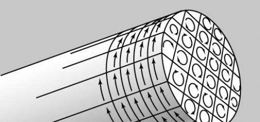
To understand the neurophysiology of the sensory and motor pathways, it is necessary to become familiar with the anatomy of the spine. The spinal cord has a structure much like a cylinder surrounded by muscle tissue. Inside the gray matter are conductive paths that control the functioning of internal organs, as well as motor functions. Associative pathways are responsible for pain and tactile sensations. Motor - for the reflex functions of the body.
As a result of trauma, malformations or diseases of the spinal cord, conduction may decrease or stop completely. This happens due to the death of nerve fibers. For a complete violation of the conduction of impulses of the spinal cord is characterized by paralysis, lack of sensitivity of the limbs. Failures in the work of internal organs begin, for which the damaged neural connection is responsible. So, with damage to the lower part of the spinal cord, urinary incontinence and spontaneous defecation are observed.
The reflex and conduction activity of the spinal cord is disturbed immediately after the onset of degenerative pathological changes. There is a death of nerve fibers that are difficult to restore. The disease progresses rapidly and a gross violation of conduction occurs. For this reason, start drug treatment needed as soon as possible.
How to restore patency in the spinal cord
The treatment of non-conductivity is primarily associated with the need to stop the death of nerve fibers, as well as to eliminate the causes that have become a catalyst for pathological changes.Medical treatment
It consists in the appointment of drugs that prevent the death of brain cells, as well as sufficient blood supply to the damaged area of the spinal cord. This takes into account the age-related features of the conductive function of the spinal cord, as well as the severity of the injury or disease.For additional stimulation of nerve cells, electrical impulse treatment is performed to help maintain muscle tone.
Surgery
The operation to restore the conduction of the spinal cord affects two main areas:- Elimination of catalysts that caused the paralysis of neural connections.
- Spinal cord stimulation to restore lost functions.
Traditional medicine for conduction disorders
Folk remedies for impaired conduction of the spinal cord, if used, should be used with extreme caution so as not to worsen the patient's condition.Particularly popular are: 
It is quite difficult to fully restore neural connections after an injury. A lot depends on quick access to medical Center and qualified neurosurgical care. The more time passes from the onset of degenerative changes, the less chance there is to restore the functionality of the spinal cord.
Morphofunctional organization of the spinal cord
The spinal cord is the most ancient part of the central nervous system of vertebrates. It is already present in the lancelet, the most primitive representative of the chordates.
The spinal cord is the caudal part of the CNS. It is placed in the spinal canal and has an unequal length in different representatives of vertebrates.
In humans, the roots of the caudal sections of the spinal cord gather in the caudal section of the spinal canal, forming the so-called cauda equina.
Spinal cord characterized by a segmental structure. The spinal cord is divided into cervical, thoracic, lumbar, sacral and coccygeal regions. Each department consists of several segments. The cervical region includes 8 segments (C 1 - C 8), the thoracic - 12 (Th 1 - Th 12), the lumbar - 5 (L 1 - L 5), the sacral - 5 (S 1 - S 5) and the coccygeal - 1- 3 (Co 1 - Co 3). Two pairs of roots depart from each segment, which correspond to one of the vertebrae and leave the spinal canal through the opening between them.
There are dorsal (rear) and ventral (anterior) roots. The dorsal roots are formed by the central axons of primary afferent neurons, whose bodies lie in the spinal ganglia.
The ventral roots are formed by axons of α- and γ-motoneurons and unmyelinated fibers of neurons of the autonomic nervous system. This distribution of afferent and efferent fibers was established independently at the beginning of the 19th century by C. Bell (1811) and F. Magendie (1822). The different distribution of functions in the anterior and posterior roots of the spinal cord is called the Bell-Magendie law. Segments of the spinal cord and vertebrae correspond to the same metamere. Nerve fibers of a pair of posterior roots go not only to their own metamere, but also above and below - to neighboring metameres. The skin area in which these sensory fibers are distributed is called the dermatome.
The number of fibers in the dorsal root is much greater than in the ventral one.
Neuronal structures of the spinal cord. The central part of the transverse section of the spinal cord is occupied by gray matter. Surrounding the gray matter is white matter. In the gray matter, anterior, posterior, and lateral horns are distinguished, and in the white matter, columns (ventral, dorsal, lateral, etc.).
The neuronal composition of the spinal cord is quite diverse. There are several types of neurons. The bodies of the neurons of the spinal ganglia are located outside the spinal cord. The axons of these neurons enter the spinal cord. The neurons of the spinal ganglia are unipolar or pseudo-unipolar neurons. In the spinal ganglia lie the bodies of somatic afferents that innervate mainly skeletal muscles. The bodies of other sensitive neurons are located in the tissue and in the intramural ganglia of the autonomic nervous system and provide sensitivity only to the internal organs. They are of two types: large - with a diameter of 60-120 microns and small - with a diameter of 14-30 microns. Large ones give myelinated fibers, and small ones - myelinated and unmyelinated. Nerve fibers of sensitive cells are classified into A-, B- and C-fibers according to the speed of conduction and diameter. Thick myelinated A fibers with a diameter of 3 to 22 microns and a conduction speed of 12 to 120 m / s are divided into subgroups: alpha fibers - from muscle receptors, beta fibers - from tactile and baroreceptors, delta fibers - from thermoreceptors, mechanoreceptors and pain receptors. To group B fibers include myelinated fibers of medium thickness with a speed of excitation of 3-14 m/s. They mainly convey the sensation of pain. To type C afferents include the majority of non-myelinated fibers with a thickness of not more than 2 microns and a conduction speed of up to 2 m / s. These are fibers that come from pain, chemo- and some mechanoreceptors.
In the gray matter of the spinal cord, the following elements are distinguished:
1) efferent neurons (motoneurons);
2) intercalary neurons;
3) neurons of the ascending tracts;
4) intraspinal fibers of sensitive afferent neurons.
motor neurons concentrated in the anterior horns, where they form specific nuclei, all of whose cells send their axons to a specific muscle. Each motor nucleus usually stretches over several segments, therefore their axons, which innervate the same muscle, leave the spinal cord as part of several ventral roots.
Interneurons are localized in the intermediate zone of gray matter. Their axons extend both within the segment and into the nearest neighboring segments. Interneurons- a heterogeneous group, the dendrites and axons of which do not leave the limits of the spinal cord. Interneurons form synaptic contacts only with other neurons, and they are the majority. Interneurons account for about 97% of all neurons. In size, they are smaller than α-motor neurons, capable of high-frequency impulses (above 1000 per second). For propriospinal intercalary neurons a characteristic property is to send long axons through several segments and terminate on motor neurons. At the same time, fibers of various descending tracts converge on these cells. Therefore, they are relay stations on the way from overlying neurons to motor neurons. A special group of intercalary neurons is formed by inhibitory neurons. These include, for example, Renshaw cells.
Ascending tract neurons are also entirely within the CNS. The bodies of these neurons are located in the gray matter of the spinal cord.
Central endings of primary afferents have their own characteristics. After entering the spinal cord, the afferent fiber usually gives rise to ascending and descending branches, which can travel considerable distances along the spinal cord. The terminal branches of one nerve afferent fiber have numerous synapses on one motor neuron. In addition, it was found that one fiber coming from the stretch receptor forms synapses with almost all motor neurons of this muscle.
Roland's gelatinous substance is located in the dorsal part of the dorsal horn.
The most accurate idea of the topography of the nerve cells of the gray matter of the spinal cord is given by dividing it into successive layers or plates, in each of which, as a rule, neurons of the same type are grouped.
According to these data, the entire gray matter of the spinal cord was divided into 10 plates (Rexed) (Fig. 2.2).
I - marginal neurons - give rise to the spinothalamic tract;
II-III - gelatinous substance;
I-IV - in general, the primary sensory area of the spinal cord (afferentation from exteroreceptors, afferentation from skin and pain sensitivity receptors);
Rice. 2.2. Division of the gray matter of the spinal cord into plates (according to Rexed)
V-VI - intercalary neurons are localized, which receive inputs from the posterior roots and descending tracts (corticospinal, rubrospinal);
VII-VIII - propriospinal intercalary neurons are located (from proprioreceptors, fibers of the vestibulo-spinal and reticulo-spinal
tracts), axons of propriospinal neurons;
IX - contains the bodies of α- and γ-motor neurons, presynaptic fibers of the primary afferents from muscle stretch receptors, the end of the fibers of the descending tracts;
X - surrounds the spinal canal and contains, along with neurons, a significant amount of glial cells and commissural fibers.
Properties of the nerve elements of the spinal cord. The human spinal cord contains approximately 13 million neurons.
α-motor neurons are large cells with long dendrites, having up to 20,000 synapses, most of which are formed by the endings of intraspinal intercalary neurons. The speed of conduction along their axon is 70-120 m/s. Rhythmic discharges with a frequency of not more than 10-20 pulses / s are characteristic, which is associated with pronounced trace hyperpolarization. These are the output neurons. They transmit signals to skeletal muscle fibers produced in the spinal cord.
γ-motor neurons are smaller cells. Their diameter is not more than 30-40 microns, they do not have direct contact with the primary afferents.
γ-motoneurons innervate intrafusal (intrafusiform) muscle fibers.
They are monosynaptically activated by the fibers of the descending tracts, which plays an important role in α-, γ-interaction. The speed of conduction along their axon is lower - 10-40 m/s. The pulse frequency is higher than that of α-motor
neurons, - 300-500 pulses / s.
In the lateral and anterior horns there are preganglionic neurons of the autonomic nervous system - their axons are sent to the ganglion cells of the sympathetic nerve chain and to the intramural ganglia of the internal organs.
The bodies of sympathetic neurons, whose axons form preganglionic fibers, are located in the intermediolateral nucleus of the spinal cord. Their axons belong to the B-fiber group. They are characterized by constant tonic impulsation. Some of these fibers are involved in maintaining vascular tone, while others provide the regulation of visceral effector structures (smooth muscles of the digestive system, glandular cells).
The bodies of parasympathetic neurons form the sacral parasympathetic nuclei. They are located in the gray matter of the sacral spinal cord. Many of them are characterized by background impulse activity, the frequency of which increases, for example, as the pressure in the bladder increases.
The spinal cord consists of two symmetrical halves, separated from each other in front by a deep median fissure, and behind by a median sulcus. The spinal cord is characterized by a segmental structure; each segment is associated with a pair of anterior (ventral) and a pair of posterior (dorsal) roots.
In the spinal cord, gray matter located in the central part and white matter lying along the periphery are distinguished.
The white matter of the spinal cord is a collection of longitudinally oriented predominantly myelinated nerve fibers. Bundles of nerve fibers that communicate between different parts of the nervous system are called tracts, or pathways, of the spinal cord.
The gray matter in cross section is butterfly-shaped and includes anterior or ventral, posterior or dorsal, and lateral or lateral horns. The gray matter contains the bodies, dendrites and (partly) axons of neurons, as well as glial cells. Basic integral part gray matter are multipolar neurons.
Cells similar in size, fine structure and functional significance lie in gray matter in groups called nuclei.
Axons of radicular cells leave the spinal cord as part of its anterior roots. The processes of internal cells end in synapses within the gray matter of the spinal cord. The axons of the beam cells pass through the white matter as separate bundles of fibers that carry nerve impulses from certain nuclei of the spinal cord to its other segments or to the corresponding parts of the brain, forming pathways. Separate areas of the gray matter of the spinal cord differ significantly from each other in the composition of neurons, nerve fibers and neuroglia.
In the posterior horns, a spongy layer, a gelatinous substance, a proper nucleus of the posterior horn, and Clark's thoracic nucleus are distinguished. Between the posterior and lateral horns, the gray matter juts into the white as strands, as a result of which its mesh-like loosening is formed, which is called the mesh formation, or reticular formation, of the spinal cord.
The posterior horns are rich in diffusely located intercalary cells. These are small multipolar associative and commissural cells, the axons of which terminate within the gray matter of the spinal cord of the same side (associative cells) or the opposite side (commissural cells).
The neurons of the spongy zone and the gelatinous substance communicate between the sensitive cells of the spinal ganglia and the motor cells of the anterior horns, closing the local reflex arcs.
Clark's nucleus neurons receive information from muscle, tendon, and joint receptors (proprioceptive sensitivity) along the thickest radicular fibers and transmit it to the cerebellum.
In the intermediate zone, there are centers of the autonomic (autonomous) nervous system - preganglionic cholinergic neurons of its sympathetic and parasympathetic divisions.
The largest neurons of the spinal cord are located in the anterior horns, which form nuclei of considerable volume. This is the same as the neurons of the nuclei of the lateral horns, radicular cells, since their neurites make up the bulk of the fibers of the anterior roots. As part of the mixed spinal nerves, they enter the periphery and form motor endings in the skeletal muscles. Thus, the nuclei of the anterior horns are motor somatic centers.
The nervous system is usually divided into several departments. According to topographic features, it is divided into central and peripheral sections, according to functional features - into somatic and vegetative sections. The central division, or central nervous system, includes the brain and spinal cord. The peripheral division, or peripheral nervous system, includes all nerves, that is, all peripheral pathways, which consist of sensory and motor nerve fibers. The somatic department, or somatic nervous system, includes cranial and spinal nerves that connect the central nervous system with organs that perceive external stimuli - with the skin and the apparatus of movement. The autonomic department, or the autonomic nervous system, provides a connection between the central nervous system and all internal organs, glands, vessels and organs, which include smooth muscle tissue. The autonomic division is divided into the sympathetic and parasympathetic parts, or the sympathetic and parasympathetic nervous system.
The central nervous system includes the brain and spinal cord. There are certain relationships between the mass of the brain and spinal cord: as the organization of the animal increases, relative mass brain compared to the spinal cord. In birds, the brain is 1.5-2.5 times larger than the spinal cord, in ungulates - 2.5-3 times, in carnivores - 3.5-5 times, in primates - 8-15 times.
Spinal cord- medulla spinalis lies in the spinal canal, occupying approximately 2/3 of its volume. In cattle and horses, its length is 1.8-2.3 m, weight 250-300 g, in pigs - 45-70 g. It looks like a cylindrical cord, somewhat flattened dorsoventrally. There is no clear boundary between the brain and spinal cord. It is believed that it passes at the level of the cranial margin of the atlas. In the spinal cord, cervical, thoracic, lumbar, sacral and caudal parts are distinguished according to their location. In the embryonic period of development, the spinal cord fills the entire spinal canal, but due to the high growth rate of the skeleton, the difference in their length becomes larger. As a result, the brain in cattle ends at the level of the 4th, in the pig - in the region of the 6th lumbar vertebra, and in the horse - in the region of the 1st segment of the sacral bone. The median dorsal sulcus (gutter) runs along the spinal cord on its dorsal side. A connective tissue dorsal septum departs from it deep into. On the sides or median sulcus are smaller dorsal lateral sulci. On the ventral side there is a deep median ventral fissure, and on the sides of it are ventral lateral grooves (troughs). At the end, the spinal cord sharply narrows, forming a brain cone, which passes into the terminal thread. It is formed by connective tissue and ends at the level of the first tail vertebrae.
There are thickenings in the cervical and lumbar parts of the spinal cord. In connection with the development of limbs in these areas, the number of neurons and nerve fibers increases. In a pig, the cervical thickening is formed by 5-8th neurosegments. Its maximum width at the level of the middle of the 6th cervical vertebra is 10 mm. Lumbar thickening falls on the 5th-7th lumbar neurosegments. In each segment, a pair of spinal nerves departs from the spinal cord with two roots - on the right and on the left. The dorsal root originates from the dorsal lateral groove, the ventral root from the ventral lateral groove. The spinal nerves leave the spinal canal through the intervertebral foramen. The section of the spinal cord between two adjacent spinal nerves is called the neurosegment. Neurosegments are of different lengths and often do not correspond in size to the length of the bone segment. As a result, the spinal nerves depart at different angles. Many of them travel some distance inside the spinal canal before leaving the intervertebral foramen of their segment. In the caudal direction, this distance increases, and from the nerves running inside the spinal canal, behind the cerebral cone, a brush is formed, as it were, called the “horse tail”.
Brain- encephalon - is placed in the cranial box and consists of several parts. In ungulates, the relative mass of the brain is 0.08-0.3% of body weight, which is 370-600 g in a horse, 220-450 g in cattle, 96-150 g in sheep and pigs. In small animals, the relative the mass of the brain is usually greater than that of large ones.
The brain of ungulates is semi-oval. In ruminants - with a wide frontal plane, with almost no protruding olfactory bulbs and noticeable extensions at the level of the temporal regions. In the pig, it is more narrowed in front, with prominent olfactory bulbs. Its length is on average 15 cm in cattle, 10 cm in sheep, and 11 cm in pigs. The brain is divided by a deep transverse fissure into a large brain lying rostrally and a rhomboid brain located caudally. Parts of the brain phylogenetically older, representing the continuation of the projection pathways of the spinal cord, are called the brain stem. It includes the medulla oblongata, the medullary bridge, the middle bridge, part of the diencephalon. Phylogenetically younger parts of the brain form the integumentary part of the brain. It includes the cerebral hemispheres and the cerebellum.
Rhomboid brain- rhombencephalon - is divided into the oblong and hindbrain and contains the fourth cerebral ventricle.
Medulla- medulla oblongata - the most rear part of the brain. Its mass is 10-11% of the mass of the brain; length in cattle - 4.5, in sheep - 3.7, in pigs - 2 cm. .
On its dorsal side there is a diamond-shaped recess - the fourth cerebral ventricle. On the ventral side there are three furrows: median and 2 lateral. Connecting caudally, they pass into the ventral median fissure of the spinal cord. Between the furrows lie 2 narrow elongated rollers - pyramids, in which bundles of motor nerve fibers pass. At the border of the medulla oblongata and spinal cord, the pyramidal tracts intersect - a cross of the pyramids is formed. In the medulla oblongata, gray matter is located inside, in the bottom of the fourth cerebral ventricle in the form of nuclei that give rise to cranial nerves (from VI to XII pairs), as well as nuclei in which impulses are switched to other parts of the brain. The white matter lies externally, predominantly ventrally, forming pathways. Motor (efferent) pathways from the brain to the spinal cord form pyramids. Sensitive pathways (afferent) from the spinal cord to the brain form / posterior legs of the cerebellum, going from the medulla oblongata to the cerebellum. In the mass of the medulla oblongata in the form of a reticular plexus lies an important coordination apparatus of the brain - the reticular formation. It integrates the structures of the brainstem and promotes their involvement in complex, multi-stage responses.
Medulla- a vital area of the central nervous system (CNS), its destruction leads to instant death. Here are the centers of respiration, heartbeat, chewing, swallowing, sucking, vomiting, chewing gum, salivation and juice secretion, vascular tone, etc.
Hind brain- metencephalon - consists of the cerebellum and the cerebral bridge.
brain bridge- pons - a massive thickening on the ventral surface of the brain, lying across the anterior part of the medulla oblongata up to 3.5 cm wide in cattle, 2.5 cm in sheep and 1.8 ohms in pigs. The bulk of the brain bridge is made up of pathways (descending and ascending) that connect the brain with the spinal cord and individual parts of the brain with each other. A large number of nerve fibers runs across the pons to the cerebellum and forms the middle cerebellar peduncles. In the bridge there are groups of nuclei, including the nuclei of the cranial nerves (V pair). From the lateral surface of the bridge departs the largest V pair of cranial nerves - trigeminal.
Cerebellum- cerebellum - located above the bridge, the medulla oblongata and the fourth cerebral ventricle, behind the quadrigemina. In front it borders on the cerebral hemispheres. Its mass is 10-11% of the mass of the brain. In sheep and pigs, its length (4-4.5 cm) is greater than its height (2.2-2.7 ohms), in cattle it approaches spherical - 5.6X6.4 cm. In the cerebellum, the middle part is distinguished - the worm and lateral parts - hemispheres of the cerebellum. The cerebellum has 3 pairs of legs. It is connected with the medulla oblongata by its posterior legs (rope bodies), the middle legs with the cerebral bridge, and the anterior (rostral) legs with the midbrain. The surface of the cerebellum is assembled into numerous folded lobules and convolutions, separated by grooves and fissures. The gray matter in the cerebellum is located above - the cerebellar cortex and in depth in the form of nuclei. The surface of the cerebellar cortex in cattle is 130 cm 2 (about 30% in relation to the cerebral cortex) with a thickness of 450-700 microns. The white matter is located under the bark and looks like a tree branch, for which it is called the tree of life.
The cerebellum is the center for coordinating voluntary movements, maintaining muscle tone, posture, and balance.
Rhomboid brain contains the fourth cerebral ventricle. Its bottom is the deepening of the medulla oblongata - the rhomboid fossa. Its walls are formed by the legs of the cerebellum, and the roof by the anterior (rostral) and posterior cerebral sails, which are the choroid plexus. The ventricle communicates rostrally with the cerebral aqueduct, caudally with the central canal of the spinal cord, and through openings in the sail with the subarachnoid space.
big brain- cerebrum - includes the terminal, diencephalon and midbrain. The telencephalon and diencephalon are combined into the forebrain.
The midbrain - mesencephalon - consists of the quadrigemina, the legs of the large brain and the cerebral aqueduct enclosed between them. Covered by large hemispheres. Its mass is 5-6% of the mass of the brain.
The quadrigemina forms the roof of the midbrain. It consists of a pair of rostral (anterior) colliculi and a pair of caudal (posterior) colliculi. The quadrigemina is the center of unconditioned reflex motor acts in response to visual and auditory stimuli. The anterior colliculi are considered the subcortical centers of the visual analyzer, the posterior colliculi are considered the subcortical centers of the auditory analyzer. In ruminants, the anterior mounds are larger than the posterior mounds; in the pig, the opposite is true.
The cerebral peduncles form the bottom of the midbrain. They look like two thick rollers lying between the visual tracts and the cerebral bridge. Separated by an interpeduncular groove.
Between the quadrigemina and the legs of the large brain in the form of a narrow tube passes the cerebral (Sylvian) aqueduct. Rostrally, it connects with the third, caudally - with the fourth cerebral ventricles. The cerebral aqueduct is surrounded by a substance of the reticular formation.
In the midbrain, the white matter is located externally and represents the conducting afferent and efferent pathways. The gray matter is located in depth in the form of nuclei. The third pair of cranial nerves departs from the brain legs.
diencephalon- diencephalon - consists of visual tubercles - thalamus, epithalamus - epithalamus, hypothalamus - hypothalamus. The diencephalon is located between the terminal.
In the midbrain, covered by the telencephalon. Its mass is 8-9% of the mass of the brain. Visual hillocks are the most massive, centrally located part of the diencephalon. Fusing between the saba, they compress the third cerebral ventricle so that it takes the form of a ring that goes around the intermediate mass of the visual tubercles. From above, the ventricle is covered with a vascular cover; communicates with the interventricular foramen with the lateral ventricles, passes aborally into the cerebral aqueduct. The white matter in the thalamus lies on top, gray - inside in the form of numerous nuclei. They serve as switching links from the underlying sections to the cortex and are connected with almost all analyzers. On the basal surface of the diencephalon is the intersection of the optic nerves - chiasm.
The epithalamus consists of several structures, including the pineal gland and the vascular tegmentum of the third cerebral ventricle (the pineal gland is an endocrine gland). It is located in the depression between the visual tubercles and the quadrigemina.
The hypothalamus is located on the basal surface of the diencephalon between the chiasm and the cerebral peduncles. Consists of several parts. Directly behind the chiasma in the form of an oval tubercle is a gray tubercle. Its downward-facing apex is elongated due to the protrusion of the wall of the third ventricle and forms a funnel on which the pituitary gland, the endocrine gland, is suspended. Behind the gray tubercle is a small rounded formation - the mastoid body. The white matter in the hypothalamus is located outside, forms the conductive afferent and efferent pathways. Gray matter - in the form of numerous nuclei, since the hypothalamus is the highest subcortical autonomic center. It contains the centers of respiration, blood and lymph circulation, temperature, sexual functions, etc.
The end brain - telencephalon - is formed by two hemispheres, separated by a deep longitudinal fissure and connected by the corpus callosum. Its mass in (cattle 250-300 g, in sheep and pigs 60-80 g, which is 62-66% of the mass of the brain. In each hemisphere, a dor-solaterally located cloak is distinguished, ventromedially - the olfactory brain, in depth - striatum and lateral ventricle.The ventricles are separated by a transparent septum.The interventricular foramen communicates with the third cerebral ventricle.
The olfactory brain consists of several parts visible on the ventral surface of the telencephalon. Rostrally, slightly protruding beyond the cloak, lie 2 olfactory bulbs. They occupy the pits of the ethmoid bone. Olfactory filaments enter them through a hole in the perforated plate of the bone, which together form the olfactory nerve. The bulbs are the primary olfactory centers. Olfactory tracts depart from them - afferent pathways. The lateral olfactory tract reaches the pear-shaped lobes, located laterally from the legs of the brain. The medial olfactory tracts reach the medial surface of the mantle. Olfactory triangles lie between the tracts. The pear-shaped lobes and olfactory triangles are the secondary olfactory centers. In the depths of the olfactory brain, at the bottom of the lateral ventricles, the remaining parts of the olfactory brain are located. They connect the olfactory brain with other parts of the brain. The striatum is located in the depths of the hemispheres and is a basal complex of nuclei, which are subcortical motor centers.
The cloak reaches its greatest development in higher mammals. It contains the highest centers of all animal life. The surface of the cloak is covered with convolutions and furrows. In cattle, its surface is 600 cm 2. The gray matter in the raincoat is located on top - this is the cerebral cortex. White matter is inside - these are pathways. The functions of different parts of the cortex are unequal, the structure is mosaic, which made it possible to distinguish several lobes in the hemispheres (frontal, parietal, temporal, occipital) and several dozen fields. The fields differ from each other in their cytoarchitectonics - the location, number and shape of cells and myeloarchitectonics - the location, number and shape of fibers.
Meninges of the brain. The spinal cord and brain are covered with hard, arachnoid and soft membranes.
The hard shell is the most superficial, thick, formed by dense connective tissue, poor in blood vessels. It fuses with the bones of the skull and vertebrae with ligaments, folds and other formations. It descends into the longitudinal gap between the hemispheres of the cerebrum in the form of a falciform ligament (sickle of the cerebrum) and separates the cerebrum from the rhomboid membranous cerebellum. Between it and the bones there is not everywhere a developed epidural space filled with loose connective and adipose tissues. This is where the veins go. From the inside, the dura mater is lined with endothelium. Between it and the arachnoid there is a subdural space filled with cerebrospinal fluid. The arachnoid membrane is formed by loose connective tissue, tender, avascular, does not enter the furrows. On both sides it is covered with endothelium and is separated by subdural and subarachnoid (subarachnoid) spaces from other membranes. Attaches to the shells with the help of ligaments, as well as vessels and nerves passing through it.
The soft shell is thin, but dense, with a large number of vessels, for which it is also called vascular. It enters all the furrows and fissures of the brain and spinal cord, as well as into the cerebral ventricles, where it forms vascular covers.
The intershell spaces, cerebral ventricles and the central spinal canal are filled with cerebrospinal fluid, which is internal environment brain and protect it from harmful effects, regulates intracranial pressure, performs a protective function. A liquid is formed. Mainly in the vascular covers of the ventricles, flows into the venous bed. Normally, its amount is constant.
Vessels of the brain and spinal cord. The spinal cord is supplied with blood by branches extending from the vertebral, intercostal, lumbar and sacral arteries. In the spinal canal they form the spinal arteries running in the sulci and the central fissure of the spinal cord. Blood enters the brain through the vertebral and internal carotid (in cattle - through the internal maxillary) arteries.