Genealogical method proposed in 1883 by F. Galton. This is a method of analyzing pedigrees (tracing the inheritance of a normal or pathological trait in a family, indicating the type of family ties between members of the pedigree). In medical genetics it is called clinical and genealogical , since pathological signs are traced and clinical research methods are applied.
The essence of the method : identification of family ties and tracing of the studied trait among close, distant, direct and indirect relatives.
Stages of the method :
1. Collecting information about relatives from the proband (a person who consulted a geneticist).
2. Drawing up a pedigree.
3. Pedigree analysis.
The method is used to establish the hereditary nature of a trait, type of inheritance, genotypes of pedigree members, and gene penetrance.
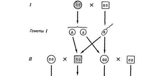
To construct genealogies, a system of symbols is used, proposed in 1931 by the English scientist Just (Fig. 17).
When constructing pedigrees, the following rules must be observed:
· it is necessary to find out the number of generations from the collected history;
· pedigree begins with the proband;
· each generation is numbered with Roman numerals on the left;
· symbols denoting individuals of the same generation are located on a horizontal line and numbered in Arabic numerals.
Pedigree analysis reveals the following types of inheritance traits: autosomal dominant; autosomal recessive; X-linked (sex-linked) dominant; X-linked (sex-linked) recessive; holandric (linked to the Y chromosome).
Autosomal dominant type of inheritance:
· A sick child is born to sick parents with a 100% probability if they are homozygous; 75% if they are heterozygous.
Figure 17. Symbolism used in compiling pedigrees
Autosomal recessive type of inheritance:
· Both men and women are affected equally.
· The probability of having a sick child from healthy parents is 25% if they are heterozygous; 0% if both or one of them is homozygous for the dominant gene.
· Often manifests itself in consanguineous marriages.
X-linked (sex-linked) dominant type of inheritance:
· Sick people occur in every generation.
· Women are more affected.
· If a father is sick, then all his daughters are sick.
· A sick child is born to sick parents with a 100% probability if the mother is homozygous; 75% if the mother is heterozygous.
· The probability of having a sick child from healthy parents is 0%.
X-chromosome (sex-linked) recessive type of inheritance:
· Patients do not occur in every generation.
· Mostly men are affected.
· The probability of having a sick boy born to healthy parents is 25%, and a sick girl is 0%.
Holandric (Y-linked) type of inheritance:
· Sick people occur in every generation.
· Only men get sick.
· If a father is sick, then all his sons are sick.
· The probability of having a sick boy from a sick father is 100%
Twin method(proposed in 1876 by F. Galton to study genetic patterns in twins.
The essence of the method : comparison of characteristics in different groups of twins based on their similarities (concordance) or differences (discordance).
Stages of the method:
1. Compiling a sample of twins from the entire population.
2. Diagnosis of zygosity of twins.
3. Establishing the relative role of heredity and environment in the formation of a trait.
To assess the role of heredity and environment in the formation and development of a trait, they use Holzinger formula:
N = ( KMB%-KDB%)/100%-KDB%
where N is the proportion of hereditary factors,
KMB% and - concordance of monozygotic twins in percentage
KDB% – concordance of dizygotic twins as a percentage
If H is greater than 0.5, then the genotype plays a large role in the formation of the trait; if H is less than 0.5, then the environment plays a large role.
Cytogenetic method is the study of karyotype using microscopy.
Stages of the method:
1. Obtaining and culturing cells (lymphocytes, fibroblasts) on artificial nutrient media.
2. Addition of phytohemagglutinin to the nutrient medium to stimulate cell division.
3. Stopping cell division at the metaphase stage by adding colchicine.
4. Treatment of cells with a hypotonic solution NaCl, as a result of which the cell membrane is destroyed and a “scattering” of chromosomes is obtained.
5. Staining of chromosomes with specific dyes.
6. Microscoping and photographing chromosomes.
7. Drawing up an idiogram and its analysis.
The method allows:
· diagnose genomic and chromosomal mutations;
Determine the genetic sex of the organism.
Biochemical methods. The cause of most hereditary monogenic diseases is metabolic defects associated with enzymopathies (disturbances in the structure of enzymes involved in metabolic reactions). At the same time, intermediate metabolic products accumulate in the body, therefore, by determining them or the activity of enzymes using biochemical methods, it is possible to diagnose hereditary metabolic diseases (gene mutations). Quantitative biochemical methods (stress tests) make it possible to identify heterozygous carriage of a pathological recessive gene.
Dermatoglyphic analysis is the study of human ridge skin (skin of the fingertips, palmar side of the hands and plantar side of the feet), where the papillary layer of the dermis is strongly pronounced.
The method is applied:
a) to establish the zygosity of twins;
b) as an express method for diagnosing the congenital component of some hereditary diseases.
Typically, with genomic pathology, a combination of certain indicators is noted: radial loops on the 4th and 5th fingers, four-digit groove, main palmar angle from 60° to 80°, etc.
Chemical methods based on high-quality color chemical reactions. Used for preliminary diagnosis of hereditary metabolic diseases. As a screening test diagnosis of phenylketonuria The method of wetting strips of paper soaked in a 10% solution of PeCl 3 or 2,4 dinitrophenyl-hydrazine with the child’s urine is used. If phenylpyruvic acid is present in the urine, a greenish color appears on the filter paper.
Determination of X- and Y-sex chromatin. Buccal epithelial cells or leukocytes are used for research. A"-chromatin is determined by staining the preparation acetorcein, and U-chromatin - when stained Acrychinipriet. These methods make it possible to identify the number of sex chromosomes in a karyotype (the number of A"-chromosomes is always one more than the number of A1-chromatin clumps, the number of Y-chromosomes is equal to the number of Y-chromatin clumps); establish the genetic sex of an individual, diagnose chromosomal diseases of sex (in combination with other methods).
Methods of prenatal (prenatal) diagnostics hereditary diseases make it possible to identify hereditary defects of the fetus in the early stages of pregnancy. With their help, it is possible to determine the disease long before the birth of the child, and if it is necessary to terminate the pregnancy.
The main indicators for prenatal diagnosis are:
· Well-established hereditary disease in the family.
· Mother's age is over 35 years, father's age is over 40 years.
· Presence of a sex-linked disease in the family.
· The presence of structural chromosome rearrangements in one of the parents (especially translocations and inversions).
· Heterozygosity of both parents for one pair of alleles in an autosomal recessive disease.
· The pregnant woman has a history of long-term work in hazardous industries or living in places with increased background radiation, etc.
· Repeated spontaneous abortions or the birth of a child with congenital malformations, diabetes mellitus, epilepsy, infections in a pregnant woman, drug therapy.
Prenatal diagnostic methods can be divided into:
1) Screening: allow us to identify women who have an increased risk of having a child with a congenital pathology or hereditary disease. The methods are widely available and relatively inexpensive. Sifting methods include:
Determination of α-fetoprotein (AFP) concentration;
Determination of the level of human chorionic gonadotropin (hCG);
Determination of the level of unbound estriol;
Detection of pregnancy-associated plasma protein A;
Isolation of fetal cells or DNA from the mother's body.
2) Non-invasive: methods of examining the fetus without surgery. Currently, these include fetal ultrasound (US). Ultrasound can be used for both screening and clarifying methods. Accumulated evidence shows that ultrasound does not harm the fetus. In some countries, ultrasound is performed on all pregnant women. This makes it possible to prevent the birth of 2-3 children with serious congenital malformations per 1,000 newborns, which is approximately 30% of all children with such a pathology.
3) Invasive: methods based on the analysis of genetic material of fetal cells or tissues. They are carried out according to strict indications. Invasive methods include:
Biopsy of the chorion and placenta (for cytogenetic, biochemical studies and DNA analysis);
Amniocentesis (sampling of fetal amniotic fluid to diagnose gene, chromosomal and genomic mutations);
Cordocentesis (taking blood from the umbilical cord for the purpose of early diagnosis of hereditary blood diseases);
Fetoscopy (introduction of a fiberoptic endoscope into the amnion cavity to examine the fetus, placenta, umbilical cord, etc.);
Nowadays, genetics is very relevant in scientific fields for research. The impetus for its development was the well-known teaching of Charles Darwin about discrete heredity, natural selection and mutational changes due to the transmission of the carrier genotype. Having begun its development at the beginning of the last century, genetics as a science has reached a wide scale, while research methods are currently one of the main areas of study of both human nature and living nature in general.
Let us consider the fundamental methods of genetic research that are currently known.
human genetics research represent the analysis and determination of typical gene structures during inheritance in pedigrees. The results and information obtained are used to prevent, prevent and identify the likelihood of the occurrence of the studied trait in the offspring - hereditary diseases. The type of inheritance can be autosomal (the manifestation of the trait is possible with equal probability in individuals of both sexes) and linked to the chromosomal sex series of the carrier.
The autosomal method, in turn, is divided into autosomal dominant inheritance (the dominant allele can be realized in both homozygous and heterozygous states) and autosomal recessive inheritance (the recessive allele can be realized only in the homozygous state). With this type of inheritance, the disease manifests itself after several generations.
Sex-linked heredity is characterized by the localization of the corresponding gene in homologous and non-homologous regions of the Y- or X-chromosomes. Based on the genotypic background, which is localized in the sex chromosomes, a hetero- or homozygous woman is determined, but men with only one X chromosome can only be hemizygous. For example, a heterozygous woman can inherit the disease to both her son and daughters.
genetics research is determined by the study of hereditary diseases transmitted as a result of gene mutations. Such methods of studying human genetics identify hereditary metabolic defects by identifying enzymes, carbohydrates and other metabolic products that remain in the extracellular fluid of the body (blood, sweat, urine, saliva, etc.).
Twin methods for studying human genetics find out the hereditary cause of the studied signs of the disease. (a full-fledged organism develops from two or more crushed parts of the zygote at an early stage of its development) have an identical genotype, which makes it possible to identify differences as a result of the external influence of the environment on the human phenotype. Fraternal twins (fertilization of two or more eggs) have the genotype of people related to each other, which makes it possible to evaluate environmental and hereditary factors in the development of a person’s genotypic background.
genetics research used in studying the morphology of chromosomes and the normality of the karyotype, which makes it possible to diagnose hereditary diseases at the chromosomal level when identifying genomic and chromosomal mutations, as well as to study the mutagenic effect of chemicals, pesticides, drugs, etc. This technique is widely used in the analysis and subsequent identification of hereditary abnormalities of the body even before the birth of a child. Prenatal diagnosis of amniotic fluid makes a diagnosis already in the first trimester of pregnancy, which makes it possible to make a decision to terminate the pregnancy.
Send your good work in the knowledge base is simple. Use the form below
Students, graduate students, young scientists who use the knowledge base in their studies and work will be very grateful to you.
Posted on http://www.allbest.ru/
inheritance somatic genealogical relative
Introduction
Genealogical method
Twin method
Cytogenetic method
Dermatoglyphic method
Biochemical method
Somatic cell hybridization
Simulation method
Immunogenetic method
Conclusion
Bibliography
Introduction
Human genetics is a branch of genetics that studies the patterns of inheritance and variability of traits in humans, closely related to anthropology and medicine. This branch is conventionally divided into anthropogenetics, which studies the heredity and variability of normal characteristics of the human body, and medical genetics. Human genetics is also associated with evolutionary theory, as it explores the specific mechanisms of human evolution and its place in nature, together with psychology, philosophy and sociology.
Genetics is one of the most rapidly developing branches of science. It is the theoretical basis of medicine and reveals the biological basis of hereditary diseases. Knowledge of the genetic nature of diseases allows you to make an accurate diagnosis in time and carry out the necessary treatment.
The study of human heredity and variability is difficult due to the inability to apply many standard approaches to genetic analysis. In particular, it is impossible to carry out directed crossing or experimentally obtain mutations. Humans are a difficult subject for genetic research also because of the large number of chromosomes; puberty occurs late; small number of descendants in each family; it is impossible to equalize living conditions for offspring.
A number of research methods are used in human genetics.
Genealogical method
The use of this method is possible in the case where direct relatives are known - the ancestors of the owner of the hereditary trait (proband) on the maternal and paternal lines in a number of generations or the descendants of the proband also in several generations. When compiling pedigrees in genetics, a certain notation system is used. After compiling the pedigree, it is analyzed in order to establish the nature of inheritance of the trait being studied.
Conventions adopted when compiling pedigrees:
1 -- man; 2 - woman; 3 -- gender is unknown; 4 - owner of the trait being studied; 5 -- heterozygous carrier of the recessive gene being studied; 6 - marriage; 7 - marriage of a man with two women; 8 - consanguineous marriage; 9 -- parents, children and their order of birth; 10 -- dizygotic twins; 11 - monozygotic twins.
Thanks to the genealogical method, the types of inheritance of many traits in humans have been determined. Thus, the autosomal dominant type inherits polydactyly (increased number of fingers), the ability to curl the tongue into a tube, brachydactyly (short fingers due to the absence of two phalanges on the fingers), freckles, early baldness, fused fingers, cleft lip, cleft palate, eye cataracts, bone fragility and many others. Albinism, red hair, susceptibility to polio, diabetes mellitus, congenital deafness and other traits are inherited as autosomal recessive.
The dominant trait is the ability to roll the tongue into a tube (1) and its recessive allele is the absence of this ability (2). 3 - pedigree for polydactyly (autosomal dominant inheritance).
A number of traits are inherited in a sex-linked manner: X-linked inheritance - hemophilia, color blindness; Y-linked - hypertrichosis of the edge of the auricle, webbed toes. There are a number of genes localized in homologous regions of the X and Y chromosomes, for example, general color blindness.
The use of the genealogical method has shown that with a related marriage, compared with an unrelated one, the likelihood of deformities, stillbirths, and early mortality in the offspring increases significantly. In consanguineous marriages, recessive genes often become homozygous, resulting in the development of certain anomalies. An example of this is the inheritance of hemophilia in the royal houses of Europe.
Inheritance of hemophilia in the royal houses of Europe: - hemophiliac; -- female carrier
Twin method
1 -- monozygotic twins; 2 - dizygotic twins.
Twins are children born at the same time. They are monozygotic (identical) and dizygotic (fraternal).
Monozygotic twins develop from one zygote (1), which at the cleavage stage is divided into two (or more) parts. Therefore, such twins are genetically identical and always of the same sex. Monozygotic twins are characterized by a high degree of similarity (concordance) for many characteristics.
Dizygotic twins develop from two or more eggs that were simultaneously ovulated and fertilized by different sperm (2). Therefore, they have different genotypes and can be of the same or different sexes. Unlike monozygotic twins, dizygotic twins are characterized by discordance - dissimilarity in many ways. Data on twin concordance for some characteristics are shown in the table.
|
Signs |
Concordance, % |
||
|
Monozygotic twins |
Dizygotic twins |
||
|
Normal |
|||
|
Blood type (AB0) |
|||
|
Eye color |
|||
|
Hair color |
|||
|
Pathological |
|||
|
Clubfoot |
|||
|
"Harelip" |
|||
|
Bronchial asthma |
|||
|
Tuberculosis |
|||
|
Epilepsy |
|||
|
Schizophrenia |
As can be seen from the table, the degree of concordance of monozygotic twins for all of the above characteristics is significantly higher than that of dizygotic twins, but it is not absolute. As a rule, discordance in monozygotic twins occurs as a result of disturbances in the intrauterine development of one of them or under the influence of the external environment, if it was different.
Thanks to the twin method, a person’s hereditary predisposition to a number of diseases was determined: schizophrenia, epilepsy, diabetes mellitus and others.
Observations of monozygotic twins provide material for elucidating the role of heredity and environment in the development of traits. Moreover, the external environment refers not only to physical environmental factors, but also to social conditions.
Cytogenetic method
Based on the study of human chromosomes in normal and pathological conditions. Normally, a human karyotype includes 46 chromosomes - 22 pairs of autosomes and two sex chromosomes. The use of this method made it possible to identify a group of diseases associated with either changes in the number of chromosomes or changes in their structure. Such diseases are called chromosomal.
The material for karyotypic analysis is most often blood lymphocytes. Blood is taken from a vein in adults, and from a finger, earlobe or heel in newborns. Lymphocytes are cultured in a special nutrient medium, which, in particular, contains added substances that “force” lymphocytes to intensively divide through mitosis. After some time, colchicine is added to the cell culture. Colchicine stops mitosis at the metaphase level. It is during metaphase that the chromosomes are most condensed. Next, the cells are transferred to glass slides, dried and stained with various dyes. Staining can be a) routine (chromosomes are stained evenly), b) differential (chromosomes acquire cross-striations, with each chromosome having an individual pattern). Routine staining makes it possible to identify genomic mutations, determine the group affiliation of a chromosome, and find out in which group the number of chromosomes has changed.
Differential staining allows you to identify chromosomal mutations, determine the chromosome by number, and find out the type of chromosomal mutation.
In cases where it is necessary to conduct a karyotypic analysis of the fetus, cells from the amniotic (amniotic fluid) fluid are taken for cultivation - a mixture of fibroblast-like and epithelial cells.
Chromosome diseases include: Klinefelter syndrome, Turner-Shereshevsky syndrome, Down syndrome, Patau syndrome, Edwards syndrome and others.
Patients with Klinefelter syndrome (47, XXY) are always men. They are characterized by underdevelopment of the gonads, degeneration of the seminiferous tubules, often mental retardation, and high growth (due to disproportionately long legs).
Turner-Shereshevsky syndrome (45, X0) is observed in women. It manifests itself in delayed puberty, underdevelopment of the gonads, amenorrhea (absence of menstruation), and infertility. Women with Turner-Shereshevsky syndrome are short, their body is disproportionate - the upper part of the body is more developed, the shoulders are wide, the pelvis is narrow - the lower limbs are shortened, the neck is short with folds, the “Mongoloid” shape of the eyes and a number of other signs.
Down syndrome is one of the most common chromosomal diseases. It develops as a result of trisomy on chromosome 21 (47; 21, 21, 21). The disease is easily diagnosed, as it has a number of characteristic signs: shortened limbs, a small skull, a flat, wide nose bridge, narrow palpebral fissures with an oblique incision, the presence of a fold in the upper eyelid, mental retardation. Disturbances in the structure of internal organs are also often observed.
Chromosomal diseases also arise as a result of changes in the chromosomes themselves. Thus, deletion of the p-arm of autosome No. 5 leads to the development of “cry of the cat” syndrome. In children with this syndrome, the structure of the larynx is disrupted, and in early childhood they have a peculiar “meowing” voice timbre. In addition, there is retardation of psychomotor development and dementia.
Most often, chromosomal diseases are the result of mutations that have occurred in the germ cells of one of the parents.
Dermatoglyphic method
In 1892 F. Galton, as one of the methods for studying humans, proposed a method for studying the skin ridge patterns of the fingers and palms, as well as the flexion palmar grooves. He established that these patterns are an individual characteristic of a person and do not change throughout life. Currently, the hereditary nature of skin patterns has been established, although the nature of inheritance has not been fully clarified. The trait is probably inherited in a polygenic manner. Dermatoglyphic studies are important in identifying twins. A study of people with chromosomal diseases revealed specific changes in them not only in the patterns of the fingers and palms, but also in the nature of the main flexion grooves on the skin of the palms. Dermatoglyphic changes in gene diseases have been less studied. These methods of human genetics are mainly used to establish paternity.
Study of prints of the skin pattern of the palms and feet. Given the existing individual differences in fingerprints, due to the developmental characteristics of the individual, several main classes are distinguished. Peculiar changes in fingerprints and palm patterns have been noted in a number of hereditary degenerative diseases of the nervous system. Characteristic of Down's disease is the monkey (four-fingered) fold, which is a line running across the entire palm in the transverse direction. Currently, the method is used mainly in forensic medicine.
Biochemical method
Hereditary diseases that are caused by gene mutations that change the structure or rate of protein synthesis are usually accompanied by disorders of carbohydrate, protein, lipid and other types of metabolism. Inherited metabolic defects can be diagnosed by determining the structure of the altered protein or its quantity, identifying defective enzymes, or detecting metabolic intermediates in extracellular body fluids (blood, urine, sweat, etc.). For example, analysis of the amino acid sequences of mutationally altered hemoglobin protein chains made it possible to identify several hereditary defects that underlie a number of diseases, including: hemoglobinosis. Thus, in sickle cell anemia in humans, abnormal hemoglobin due to mutation differs from normal by replacing only one amino acid (glutamic acid with valine).
In healthcare practice, in addition to identifying homozygous carriers of mutant genes, there are methods for identifying heterozygous carriers of some recessive genes, which is especially important in medical genetic counseling. Thus, in phenotypically normal heterozygotes for phenylketonuria (a recessive mutant gene; in homozygotes, the metabolism of the amino acid phenylalanine is disrupted, which leads to mental retardation), after taking phenylalanine, an increased level of phenylalanine in the blood is detected. In hemophilia, heterozygous carriage of a mutant gene can be established by determining the activity of the enzyme altered as a result of the mutation.
Population statistical method
This is a method for studying the distribution of hereditary traits (hereditary diseases) in populations. An essential point when using this method is the statistical processing of the data obtained. A population is understood as a collection of individuals of the same species, living for a long time in a certain territory, freely interbreeding with each other, having a common origin, a certain genetic structure and, to one degree or another, isolated from other such collections of individuals of a given species. A population is not only a form of existence of a species, but also a unit of evolution, since the microevolutionary processes that culminate in the formation of a species are based on genetic transformations in populations.
A special branch of genetics deals with the study of the genetic structure of populations - population genetics.
There are three types of populations in humans:
1) panmictic,
3) isolates that differ from each other in numbers, frequency of intragroup marriages, proportion of immigrants, and population growth. The population of a large city corresponds to a panmictic population.
The genetic characteristics of any population include the following indicators:
1) gene pool (the totality of genotypes of all individuals in a population),
2) gene frequencies,
3) genotype frequencies,
4) frequencies of phenotypes, marriage system,
5) factors that change gene frequencies.
To determine the frequency of occurrence of certain genes and genotypes, the Hardy-Weinberg law is used.
Hardy-Weinberg Law
In an ideal population, from generation to generation, a strictly defined ratio of the frequencies of dominant and recessive genes is maintained (1), as well as the ratio of the frequencies of genotypic classes of individuals (2).
p + q = 1, (1)
p2 + 2pq + q2 = 1, (2)
Where p -- frequency occurrence dominant gene A; q -- frequency occurrence recessive gene A; p2 -- frequency occurrence homozygotes By dominant AA; 2pq -- frequency occurrence heterozygotes Aa; q2 -- frequency occurrence homozygotes By recessive ah.
The ideal population is a sufficiently large, panmictic (panmixia - free crossing) population, in which there is no mutation process, natural selection and other factors that disturb the balance of genes. It is clear that ideal populations do not exist in nature; in real populations, the Hardy-Weinberg law is used with amendments.
The Hardy-Weinberg law, in particular, is used to approximate the number of carriers of recessive genes for hereditary diseases. For example, phenylketonuria is known to occur at a frequency of 1:10,000 in this population. Phenylketonuria is inherited in an autosomal recessive manner, therefore, patients with phenylketonuria have the aa genotype, that is, q2 = 0.0001. Hence: q = 0.01; p = 1 - 0.01 = 0.99. Carriers of the recessive gene have the genotype Aa, that is, they are heterozygotes. The frequency of occurrence of heterozygotes (2pq) is 2 · 0.99 · 0.01? 0.02. Conclusion: in this population, about 2% of the population are carriers of the phenylketonuria gene. At the same time, you can calculate the frequency of occurrence of homozygotes by dominant (AA): p2 = 0.992, slightly less than 98%.
A change in the balance of genotypes and alleles in a panmictic population occurs under the influence of constantly acting factors, which include: mutation process, population waves, isolation, natural selection, genetic drift, emigration, immigration, inbreeding. It is thanks to these phenomena that an elementary evolutionary phenomenon arises - a change in the genetic composition of the population, which is the initial stage of the process of speciation.
Comparative genetic method
One of the methods of human genetics is the comparative genetic method, or the biomodeling method, the theoretical basis of which is the law of homological series of hereditary variability.
This method is based on the use of laboratory animals with which experiments can be carried out, including targeted crossbreeding, administration of chemicals, irradiation and dissection at the required time. Since mammals have a large number of “common” (identical in origin) genes, information obtained in experiments on animals can be transferred to humans.
The comparative genetic method is of particular importance in medical genetics, where it makes it possible to determine the genetic causes and mechanisms of development of hereditary human diseases that are also found in animals, and to develop methods of treating them using animals. Models of human diseases such as hemophilia (low blood clotting), phenylketonuria (increased concentration of phenylpyruvic acid in the urine and blood plasma, leading to dementia), various anemia (anemia), atherosclerosis (deposition of lipids on the walls of blood vessels), hypertension (high blood pressure), obesity, breast cancer, etc.
Somatic cell hybridization
Using these methods, the heredity and variability of somatic cells are studied, which compensates for the impossibility of applying hybridological analysis to humans. These methods, based on the reproduction of these cells in artificial conditions, analyze genetic processes in individual cells of the body, and, thanks to the usefulness of the genetic material, use them to study the genetic patterns of the whole organism.
Hybrid cells containing 2 complete genomes usually “lose” chromosomes of preferably one of the species when dividing. Thus, it is possible to obtain cells with the desired set of chromosomes, which makes it possible to study the linkage of genes and their localization on certain chromosomes.
Thanks to the methods of somatic cell genetics, it is possible to study the mechanisms of primary action and interaction of genes, the regulation of gene activity. The development of these methods has determined the possibility of accurate diagnosis of hereditary diseases in the prenatal period.
Simulation method
Studies human diseases in animals that can suffer from these diseases. It is based on Vavilov’s law on homologous series of hereditary variability, for example, sex-linked hemophilia can be studied in dogs, epilepsy in rabbits, diabetes mellitus, muscular dystrophy in rats, cleft lip and palate in mice.
Models in biology are used to simulate biological structures, functions and processes at different levels of organization of living things: molecular, subcellular, cellular, organ-systemic, organismal and population-biocenotic. It is also possible to model various biological phenomena, as well as the living conditions of individuals, populations and ecosystems.
In biology, mainly three types of models are used: biological, physicochemical and mathematical (logical-mathematical).
Biological models reproduce in laboratory animals certain conditions or diseases found in humans or animals. This allows us to study experimentally the mechanisms of occurrence of a given condition or disease, its course and outcome, and influence its course. Examples of such models are artificially induced genetic disorders, infectious processes, intoxication, reproduction of hypertensive and hypoxic conditions, malignant neoplasms, hyperfunction or hypofunction of certain organs, as well as neuroses and emotional states. To create a biological model, various methods are used to influence the genetic apparatus, infection with microbes, introduction of toxins, removal of individual organs or introduction of their waste products (for example, hormones), various effects on the central and peripheral nervous system, exclusion of certain substances from food, placement into an artificially created habitat and many other ways. Biological models are widely used in genetics, physiology, and pharmacology.
Immunogenetic method
The immunological (serological) method includes the study of blood serum, as well as other biological substrates to identify antibodies and antigens.
There are serological reactions and immunological methods using physical and chemical labels. Serological reactions are based on the interaction of antibodies with antigens and registration of accompanying phenomena (agglutination, precipitation, lysis). In immunological methods, physical and chemical labels are used that are included in the formed antigen-antibody complex, allowing the formation of this complex to be recorded.
Classical serodiagnosis is based on the determination of antibodies to an identified or suspected pathogen. A positive reaction result indicates the presence of antibodies to pathogen antigens in the blood serum being tested; a negative result indicates the absence of such.
Serological reactions are semi-quantitative and allow one to determine the antibody titer, i.e. the maximum dilution of the test serum in which a positive result is still observed.
The detection of antibodies to the causative agent of a number of infectious diseases in the tested blood serum is not enough to make a diagnosis, since it may reflect the presence of post-infectious or post-vaccination immunity. That is why paired sera are examined - taken in the first days of the disease and after 7-10 days. In this case, the increase in antibody titer is assessed. A diagnostically significant increase in antibody titer in the test blood serum relative to the initial level is 4 times or more. This phenomenon is called seroconversion.
In exotic infectious diseases, as well as in hepatitis, HIV infection and some other diseases, the very fact of detecting antibodies indicates that the patient is infected and has diagnostic value.
Conclusion
Advances in the development of human genetics have made it possible to prevent and treat hereditary diseases. One of the effective methods of their prevention is medical genetic counseling with prediction of the risk of the appearance of the patient in the offspring of persons suffering from this disease or having a sick relative.
Advances in human biochemical genetics have revealed the root cause (molecular mechanism) of many hereditarily determined defects and metabolic abnormalities, which has contributed to the development of rapid diagnostic methods that allow patients to be quickly and early identified, and the treatment of many previously incurable hereditary diseases. Most often, treatment consists of introducing into the body substances that are not formed in it due to a genetic defect, or in preparing special diets from which substances that have a toxic effect on the body as a result of a hereditary inability to break them down are eliminated.
Many genetic defects are corrected with timely surgical intervention or pedagogical correction.
Practical measures aimed at maintaining human hereditary health and protecting the gene pool of humanity are carried out through a system of medical genetic consultations. Their main goal is to inform interested parties about the likelihood of the risk of the appearance of patients in the offspring. Medical genetic activities also include the promotion of genetic knowledge among the population, as this promotes a more responsible approach to childbearing. Medical genetic consultation refrains from coercive or encouraging measures in matters of childbirth or marriage, taking on only the function of information. Of great importance is a system of measures aimed at creating the best conditions for the manifestation of positive hereditary inclinations and preventing the harmful effects of the environment on human heredity.
Human genetics represents the natural scientific basis for the fight against racism, convincingly showing that races are forms of human adaptation to specific environmental conditions (climatic and other), that they differ from each other not by the presence of “good” or “bad” genes, but by frequency the spread of common genes common to all races. Human genetics shows that all races are equivalent (but not the same) from a biological point of view and have equal opportunities for development, determined not by genetic, but by socio-historical conditions. The statement of biological hereditary differences between individuals or races cannot serve as a basis for any moral, legal or social conclusions that infringe on the rights of these individuals or races.
Bibliography
1. Fundamentals of ecology./ ed. Obukhova V. L. and Sapunova V. B. S.-Pb: Special literature, 1998.
2. Ruzavin G.I. Concepts of modern natural science. M.: Unity, 2010.
3. Sheppard F. M. Natural selection and heredity. M.: Education, 2009.
4. . http://schools.keldysh.ru/sch1952/Pages/Timokhina04/Biolog/18.htm.
5. . http://www.licey.net/bio/biology/lection22.
6. . https://sites.google.com/site/biologiasch88/u/sipicyna-a-i-orlova-t/metody-genetiki.
Posted on Allbest.ru
...Similar documents
The essence of the genealogical method and its application in human genetics. Peculiarities of inheritance of various traits. Hypothesis and progress of pedigree research. Genetic patterns of inheritance of human traits and comparison of results with the hypothesis.
practical work, added 05/20/2009
The patterns of inheritance and variability of traits in humans are the subject of the study of genetics. Characteristics of the main research methods. Method of compiling pedigrees (genealogical). Population, twin, cytogenetic, biochemical methods.
presentation, added 04/11/2015
Disclosure of the essence of gynealogical, twin, cytogenetic and population methods for studying hereditary characteristics. Chromosomal analysis of the human genetic code, major genetic diseases. Albinism, Down and Marfan syndromes.
presentation, added 09.09.2014
Study of an experiment on the Drosophila fly to study the heredity and variability of species. Reprogramming of somatic cells. The principle of using induced pluripotent stem cells. Method of transferring the nucleus of a somatic cell into an oocyte.
course work, added 04/02/2015
The main stages of development, tasks and sections of genetics, its influence on other branches of biology. Characteristics of the main methods of studying heredity: genealogical, twin, biochemical, cytogenetic (karyotypic) and population.
abstract, added 03/10/2012
Application of cell technologies in plant breeding. Use of in vitro methods in remote hybridization. Work on cultivating callus in order to obtain new breeding material. Hybridization of somatic cells and its main results.
abstract, added 08/10/2009
Development of the recombinant DNA method. Analysis of the inheritance of family diseases and the study of genetic linkage in humans in cases where complications arise: genetic heterogeneity and phenocopies. Genetic linkage map of the human genome.
tutorial, added 08/11/2009
The essence of the genealogical method. Diagnosis of risk level in genetic situations. Similarities between identical and fraternal twins. Study of the human chromosome set. Mutations that occur in the germ cells of one of the parents during meiosis.
presentation, added 11/04/2015
Human genome. Genetic products. Determination of paternity using DNA diagnostics. Fingerprint identification of a person. Histological and cytological research methods in forensic medicine. Century of biology and genetics.
abstract, added 04/18/2004
Features and methods of studying human genetics. Inheritance of individual human characteristics. Autosomal dominant type of inheritance. Sex-linked traits. Conventions adopted for compiling pedigrees. Chromosomal diseases.
A person as an object of genetic research has almost no advantages over other objects.
On the contrary, there are many obstacles that make it difficult to study its genetics: 1) the impossibility of random crossing in an experiment; 2) late onset of puberty; 3) a small number of descendants in each family; 4) the impossibility of equalizing living conditions for offspring; 5) lack of accurate registration of the manifestation of hereditary properties in families and the absence of homozygous lines; 6) a large number of chromosomes; 7) and the most important difficulty in studying human genetics in a capitalist society is social inequality, which makes it difficult to realize a person’s hereditary potential.
Despite these difficulties, genetics has developed some methods that make it possible to study heredity and inheritance in humans step by step. There are several research methods: genealogical, cytogenetic, twin, ontogenetic and population.
It should be borne in mind that any trait, regardless of whether it is a wild-type trait, i.e., normal, or associated with a disease, can serve as a model for the study of heredity. Protecting a person from hereditary diseases or damage to his heredity is just as important as finding out the inheritance of the norm. Currently, genetic methods have been developed mainly in relation to morphological characteristics that are genetically determined quite clearly (brachydactyly, albinism, color blindness, mottled skin and hair, etc.).
Genetic research of mental properties still remains problematic, since elementary criteria for a trait in the genetic sense have not been found for them. Almost all the signs of human mental and creative activity are so complex and complex, and are also strongly conditioned by external, including social, factors that a genetic analysis of these properties is still difficult to carry out, although their hereditary conditionality is beyond doubt.
It can be said that the vast majority of traits characterizing the species Homo sapiens can be studied as quantitative and complex physiological traits, that is, traits that do not exhibit a discrete nature in ontogenesis. These traits are controlled by the genotype system (polygenic). And until this system is solved, at least using the example of simply organized organisms, the problem of behavioral traits remains inaccessible to genetic analysis. On the contrary, mutant characters that go beyond the boundaries of the characteristics of species characters serve as good genetic models for the study of heredity and inheritance under normal conditions.
Discrete mutant traits cannot be viewed as merely pathological traits that supposedly have no adaptive significance. It is possible that the very appearance of a person with developed hemispheres of the cerebral cortex, a vertical body position, and discrete speech signaling is a consequence of major mutations. This is strongly supported by
a short period of human evolution, during which small mutations could hardly accumulate in such quantities and give such a significant evolutionary effect. A reasonable person is as “unusual” to nature as a domestic chicken that lays 365 eggs per year instead of 10-15, or a record-breaking cow that produces 16 thousand kg of milk per year instead of 600-700 kg.
The division of traits into normal and mutant in relation to humans and animals is necessary for understanding human evolution and pathological phenomena.
The set of species characteristics of humans and animals is determined by the genotype system, which has developed under the influence of all selection factors in the process of evolution. Mutations that are in a heterozygous state in humans are apparently as necessary as in animals to maintain them in the population.
The most dangerous thing in the development of scientific methods for studying animals and humans, especially their abilities, is the anthropomorphic moment, i.e., wishful thinking as reality.
Genealogical method
Analysis of human inheritance based on the compilation of a pedigree - genealogy was proposed by F. Galton.
Genealogical method is the study of the inheritance of human properties through pedigrees (pedigree). This method is applicable if direct relatives are known - the ancestors of the owner of a hereditary trait (proband) on the maternal and paternal lines in a number of generations and there is a sufficient number of descendants in each generation, or in the case when there is data on a sufficient number of different families to identify similarities pedigrees. Data on a set of similar pedigrees are subjected to statistical processing.
The most widespread system for designating human pedigrees was proposed by G. Just in 1931.
Based on a large number of analyzed families, pedigrees are compiled and mathematical calculations are made according to the type of inheritance of a particular trait - dominant or recessive, frequent and infrequent mutations, sex-linked or not, etc. Here we will not touch on the application of the mathematical method To this analysis, we only note that this entire formal analysis is based on elementary genetic laws of inheritance.
Pedigree patterns of inheritance of a dominant autosomal gene that determines a trait, for example, a disease (chondrodystrophic dwarfism, epidermolysis bullosa - the ability of the skin to form large blisters with minor injuries, retinoblastoma, etc.), or a morphological defect, for example short-fingered feet (brachydactyly - the absence of two distal phalanges in the fingers).

The inheritance of traits determined by recessive genes (recessive inheritance) is analyzed somewhat more complexly when drawing up pedigree charts.
For example, two in a family, the appearance of two sick children is equal to the product of the probabilities, i.e. 0.25 X 0.25, i.e. 6.25%.

Frequently occurring recessive autosomal genes, provided that their carriers (aa) are able to marry and produce offspring, will be in high concentration in the population. In this case, marriages aa X Aa become very probable, in the offspring from which the inheritance of this trait will imitate inheritance according to the dominant type 1:1. However, knowing the type of inheritance and the manifestation of these and other genes, even in the case of small families, but with a sufficient number of such families, it is possible to establish the true nature of inheritance.
The inheritance of genes that are completely sex-linked, i.e., located in non-homologous segments, and partially sex-linked, localized in homologous segments of the X- and Y-chromosomes, obeys the laws established for sex chromosomes. For dominant and recessive genes, this inheritance will be determined differently depending on where the gene is localized - in a homologous or non-homologous segment of the X and Y chromosomes and how it is transmitted. Thus, the dominant gene that causes webbed fingers, located in the non-homologous segment of the Y chromosome, is inherited from fathers and appears only in men.
For partially sex-linked dominant genes located in homologous segments of sex chromosomes, the analysis is somewhat more difficult, but is also possible. An example of a sex-linked inheritance of a recessive trait is the inheritance of hemophilia. There is discontinuity in the transmission of this trait across generations; affected men are the offspring of healthy mothers who were heterozygotes for this gene; Women with hemophilia may be the offspring of a sick father and a sick or healthy mother.

About 50 sex-linked recessive genes have been found in humans. Interestingly, about half of them cause eye disease. It has been known since ancient times that the degree of transmission of hereditary characteristics in related (inbreeding) and unrelated marriages (outbreeding) is different. After genetics has established patterns of more frequent manifestation of recessive genes during inbreeding, there is no need to prove at length the harm of consanguineous marriages. The higher the inbreeding coefficient, the greater the likelihood of hereditary diseases occurring over generations. In different countries, among different peoples and classes of society, as well as in different eras, consanguineous marriages (between first and second cousins) occur with different frequencies. For example, in villages on the Fiji islands the number of consanguineous marriages reaches 29.7%, in some castes of India - 12.9, in Japan (Nagasaki) - 5.03, in Holland - 0.13-0.159, in Portugal - 1 .40, in the USA (Baltimore) - 0.05%, etc. The percentage of consanguineous marriages fluctuates in certain areas of the same country depending on the way of life.
The harmfulness of consanguineous marriages is little noticeable in individual pedigrees, but in a comparative statistical analysis of diseases and mortality, it becomes completely obvious.

A striking example of identifying a recessive gene in a consanguineous marriage.

In this pedigree, kinship is maintained through the marriage of sibs (brothers - sisters) of varying degrees of kinship. From two consanguineous marriages (fourth cousins), 4 out of 8 children appeared in one family, and in the other - 2 out of 5 children suffering from hereditary amaurotic idiocy. K. Stern suggests that one of the two common ancestors of these lines passed on this recessive gene through three generations to each of the four parents.
Sometimes the disease and mortality of children from consanguineous marriages exceed by 20-30% those from unrelated marriages. Obviously, the reason for the phenomenon under consideration is genetic, namely: a high probability of the manifestation of hereditary diseases and mortality as a result of homozygotization of recessive genes that determine physiological deficiencies and mortality (lethal and semi-lethal genes).
So, the genealogical method is a very valuable method, but its importance in research is greater, the more accurately and deeply the genealogies are compiled. As civilization grows and pedigrees are recorded more accurately, the role of this method in human genetics will increase.
Twin method
Gemini refers to the offspring consisting of simultaneously born individuals in single-bearing animals (humans, horses, cattle, sheep, etc.).
Twins can be identical or fraternal.
Identical, or identical, twins(OB) develop from one egg fertilized by one sperm, when two or more embryos arise from a zygote instead of one (polyembryony). Due to the fact that the mitotic division of the zygote produces two equally hereditary blastomeres, identical twins, no matter how many of them develop, must be hereditarily identical and of the same sex. This phenomenon is an example of asexual, or more precisely, vegetative reproduction of animals.
Fraternal twins(RB) develop from simultaneously ovulated different eggs fertilized by different sperm. And since different eggs and sperm can carry different combinations of genes, fraternal twins can be hereditarily as different as children of the same couple born at different times. Fraternal twins can be of the same sex (RBo) or different sexes (RBr).
More often in the literature, instead of the term “fraternal twins” (RB), the term “fraternal twins” (DT) is used, since twins are more common. However, the term “fraternal twins” better emphasizes the difference between OB and RB; Identical twins are also more likely to be born twins.
Judging by the data obtained on mammals, there may be several hypotheses to explain the formation of OB in humans:
- divergence of blastomeres during the first division of the zygote and separate development of the embryo from these blastomeres;
- separation of a group of cells at the blastocyst stage (before gastrulation);
- separation of embryos at the early stage of gastrulation. The most likely route is the second one.

The number of twins in one birth in a person varies: twins are most common, triplets are less common, quadruplets are even less common, and quintuplets are very rare. According to I.I. Kanaev, over the past 150 years, four cases of quintuplet births have been identified in the United States, and two cases in Canada. The fact of the birth of OB - five girls who survived to adulthood - is known in the family of the Canadian farmer Dionne (1934). It is calculated that quintuplets are born once in 54,700,816 births, gears - in 4,712 million births, septuplets are known only as an exception. The average twin birth rate is 1%, with fluctuations ranging from 0.5-1.5%. Twins are less viable, and therefore their number at birth is less than at conception, and in adulthood less than at birth.
The calculation of the frequency of OB in relation to RB is done based on the theoretical ratio of same-sex and opposite-sex pairs of RB at the birth of twins: 25%♀♀ + 50%♀♂ + 25%♂♂ subtracting the number of pairs of different sexes from the total number of all pairs of the same sex (male and female) will give a difference equal to the number of OB pairs, which on average ranges from 21 to 33.4% of all twins.
To use twins in genetic studies, it is very important to accurately diagnose the OB type and the RB type. Diagnosis is made based on the following criteria:
- OBs are necessarily of the same sex, RBs can be either the same sex or different sexes;
- OB, as a rule, have one common chorion, RB - different chorions;
- reciprocal tissue transplantation in OB is as successful as autotransplantation; in RB it is impossible;
- the presence of similarity (concordance) in OB and dissimilarity (discordance) in RB for many characteristics.
For diagnosis, one should select characteristics that are clearly inherited and least susceptible to change under the influence of environmental factors; These signs include blood types, pigmentation of the eyes, skin and hair, skin relief (prints of fingertips, palms, feet, etc.). If twins are found to be different based on one or two of these characteristics, then they are usually RB.
All doubtful cases of diagnosing twins can be caused either by a developmental disorder of one of the OB partners, or by the similarity of the parents in a number of ways. However, the latter is extremely rare. It should be noted that disruption of the development of one of the OB partners is usually explained by the unequal effect of factors in intrauterine life and the occurrence of somatic mutations in the early stages of embryonic development, before the formation of organs. Various gene and chromosomal rearrangements, monosomy and other mutations that occur in one of the partners can cause significant differences in the OB phenotype. Therefore, it is necessary to take into account the possibility of somatic mutations in OB in early embryogenesis.
According to the generalizations of I. I. Kanaev, set out in his excellent monograph, the essence of the twin method in genetics comes down to the following provisions:
1) the OB pair has an identical combination, the RB pair has different combinations of parent genotypes;
2) for both partners of one pair of OBs, the external environment may be the same, but for the other, different. If OB partners experience different influences over the course of their lives, this will lead to within-pair differences. Hence, pairs can have intra-pair identical and intra-pair different environments.
Comparing OB with the same environment with OB with different environments opens up the possibility of judging the role of environmental influences on intrapair differences between twins throughout life. Comparison of OB with the same environment and RB with the same environment makes it possible to clarify the role of the hereditary factor. This kind of study is carried out on a large sample and processed statistically.
Based on the difference in the genetic origin of OB and RB, it follows that if OB and RB have no differences in the same characteristics, then it is obvious that these differences in traits in the latter are due to hereditary factors. If intrapair differences in the same characteristics occur in one and the other type of twin, then it is obvious that they can be caused by environmental factors. From the data on discordance in OB and RB for a number of morphological characteristics, it is clear that intrapair differences in RB occur many times more often than in OB.

Some data from S. Reed are presented regarding the comparative frequency of pathology in the second partner in the case of illness in one of the twins.

The percentage shows the frequency of concordance of diseases in two types of twins; it shows that if one partner fell ill with one of the indicated diseases, then the probability of the second partner getting sick in OB is much higher than in RB. V.P. Efroimson, analyzing data on the frequency of contorted couples, quite correctly points out that a high hereditary predisposition of OB to diseases manifests itself in the presence of a provoking factor; without it, this percentage will be significantly lower.
The twin method makes it possible to determine with the greatest accuracy a person’s hereditary predisposition to a number of diseases and properties. With other methods it is very difficult or almost impossible to study many infectious and tumor diseases, inflammations of the skin and various organs, as well as the characteristics of normal human nervous activity.
When using the twin method, it is necessary to take into account the conditions of joint and separate upbringing in the lives of partners, the social conditions in which they find themselves, etc. Nevertheless, the twin method makes it possible to most accurately determine the heritability coefficient of various traits, as well as judge the heterogeneity of the population based on the genes being studied and identifying the role of the environment in determining the variability of the studied traits.
Cytogenetic method
Cytogenetic method In human genetics, cytological analysis of a person’s karyotype in normal and pathological conditions is usually called.
It is more correct to call this method cytological rather than cytogenetic, since genetic analysis by crossing in humans is excluded, and carriers of chromosomal abnormalities, if they survive, are usually infertile. However, occasionally, for some chromosomal disorders, it is possible to combine the cytological method with the genealogical one and establish a connection between the phenotypic effect and a certain type of chromosomal changes. Due to these circumstances, it is possible to retain the term “cytogenetic method” accepted in the literature in the study of human genetics. In those cases where such parallelism is not being studied, the use of this term is unauthorized.
The cytogenetic method is used to study various types of heteroploidy and chromosomal rearrangements in human somatic tissues, causing various phenotypic deviations from the norm.
This method is most often used in tissue culture. It allows you to take into account major chromosome abnormalities that occur in both germ and somatic cells. It turned out that in humans, as well as in animals, trisomics and monosomics quite often occur in different pairs of chromosomes due to non-disjunction of autosomes and sex chromosomes in meiosis. Trisomy and monosomy of sex chromosomes in humans are detected based on sex chromatin analysis.
During the relatively long individual development of a person, chromosomal abnormalities (chromosomal rearrangements, as well as changes in the number of chromosomes) accumulate in the cells of various tissues. Body tissues represent diverse populations of genetically different cells, in which the concentration of cells with pathological nuclei increases with age. In this case, the cytogenetic method makes it possible to study tissue aging based on the study of cell structures in the age-related dynamics of the “population” of somatic and generative tissues.
Since the frequency of occurrence of chromosomal abnormalities depends on the influence of various mutagens on the body (ionization, chemical agents - pharmacological drugs, gas composition of the environment, etc.), the cytogenetic method makes it possible to determine the mutagenic effect of environmental factors on humans.
The use of the cytogenetic method has especially expanded in connection with the discovery of the causes of a number of physical and mental diseases - the so-called chromosomal diseases.
There are several human diseases, for example Klinefelter's disease, Shereshevsky-Turner's disease, Down's disease, etc., the causes of which remained unknown for a long time until chromosomal abnormalities were discovered in such patients by cytological methods.
Sick men with Klinefelter syndrome are characterized by underdevelopment of the gonads, degeneration of the seminiferous tubules, mental retardation, disproportionate growth of the limbs, etc. Shereshevsky-Turner syndrome occurs in women. It manifests itself in delayed puberty, underdevelopment of the gonads, absence of menstruation, infertility, short stature and other pathological signs.
It turned out that both of these syndromes in offspring are a consequence of nondisjunction of sex chromosomes during the formation of parental gametes. Due to the non-disjunction of the X chromosomes in the female homogametic sex, during the process of meiosis, gametes can arise with two X chromosomes, i.e. XX + 22 autosomes, and without X chromosomes, i.e. 0 + 22; in the male (heterogametic) sex, the gametes are XY + 22 and 0 + 22, respectively. In the case of fertilization of such eggs by normal sperm (X + 22 or Y + 22), the formation of the following classes of zygotes is possible: XXX + 44, 0X + 44 and XXY + 44, 0Y + 44.
It follows from this that the number of chromosomes in zygotes of different origins can vary from 47 to 45, and individuals 0Y + 44 obviously do not survive, since they have never been found. The chromosome set XXY + 44 is inherent in a man with Klinefelter syndrome (male intersex), chromosome sets X0 + 44 and XXX + 44 are found in women with Shereshevsky-Turner syndrome.
Upon further analysis of patients with different syndromes, it turned out that due to nondisjunction of sex chromosomes, various types of chromosomal abnormalities, in particular polysomy, can occur. There are, for example, men with the following sets of chromosomes: XX Y, XXX Y, XXXX Y, and women - XXX, XXXX.
The peculiarity of the role of sex chromosomes in the determination of sex in humans in the case of their nondisjunction, in contrast to Drosophila, is manifested in the fact that the set of chromosomes XX Y always determines the male sex, and the set X0 - the female. Moreover, an increase in the number of X chromosomes in combination with one Y chromosome does not change the definition of male sex, but only enhances Klinefelter syndrome. Trisomy, or polysomy on the X chromosome, also often causes diseases similar to Shereshevsky-Turner syndrome in women.
Diseases caused by a violation of the normal number of sex chromosomes are diagnosed using a cytological method - sex chromatin analysis. In cases where the tissues of men have a normal set of sex chromosomes - XY, sex chromatin is not detected in the cells. In normal women - XX - it is found in the form of one body. With polysomy on X chromosomes in women and men, the number of sex chromatin bodies is always one less than the number of X chromosomes, i.e. n x = n X - 1. Thus, in the cells of men with Klinefelter syndrome with a set of XX Y there is one body sex chromatin, when dialing XXXY - two, when dialing XXXXY - three; in women with Shereshevsky-Turner syndrome, respectively: X0 - no body, XXX - two bodies, XXXX - three bodies of sex chromatin, etc. It is assumed that in each such zygote only one of the X chromosomes is genetically active. The remaining chromosomes enter the heteropyknotic state in the form of sex chromatin.
The reasons for this pattern have not been clarified, but it is assumed that it is associated with the leveling of the action of sex chromosome genes in the hetero- and homogametic sex.
As we know, chromosome nondisjunction can occur not only in meiosis, but also in somatic cells throughout the entire embryogenesis of an animal, starting from the first egg cleavage. Due to the latter, among people, when the divergence of sex chromosomes is disrupted, sick female mosaics and male mosaics may appear. For example, mosaics of the following types are described: double: X0/XX, X0/XXX and X0/XY, X0/XYY, triple: X0/XX/XXX, XX/X0/XY, as well as quadruple mosaics, when somatic cells of one humans contain four different sets of chromosomes.
In addition to the considered type of diseases caused by changes in the number of sex chromosomes in the zygote, chromosomal diseases can be caused by autosomal nondisjunction and various kinds of chromosomal rearrangements (translocations, deletions). For example, in children with congenital idiocy - Down's disease, accompanied by short stature, a wide round face, closely spaced narrow palpebral fissures and a half-open mouth, trisomy 21 was discovered. It has been established that the incidence of Down syndrome in newborns depends on the age of the mothers.
A wide variety of diseases are associated with congenital chromosomal abnormalities. Therefore, the cytogenetic method is becoming important in the etiology of human diseases.
Population method
Population method allows you to study the distribution of individual genes or chromosomal abnormalities in human populations.
The population method is based on mathematical methods. To analyze the genetic structure of a population, it is necessary to examine a large sample, which must be representative - objectively reflect the entire general population, i.e. the entire population as a whole. In the surveyed sample, the distribution of individuals into corresponding clearly defined phenotypic classes is established, the differences between which are hereditarily determined. Then, based on the found phenotypic frequencies, gene frequencies are determined.
Based on knowledge of gene frequencies, it is possible to describe the analyzed population in accordance with the Hardy-Weinberg formula and predict in advance the likely nature of segregation in the offspring of individuals belonging to certain phenotypic classes. The study of gene frequencies is important for assessing the consequences of consanguineous marriages, as well as for elucidating the genetic history of the human population as a whole.
The frequency of distribution in populations of different anomalies turns out to be different; Moreover, the overwhelming number of corresponding recessive alleles are presented in the heterozygous state.
Thus, approximately every hundredth inhabitant of Europe is heterozygous for the amaurotic idiocy gene (Spilmeier-Vogt disease), while only 25 out of 1 million people who are homozygous develop this disease in adolescence. Albinos in European countries occur with a frequency of 1 in 20,000, although the heterozygous state of this allele is inherent in every seventieth inhabitant.
The situation is somewhat different in the case of anomalies inherited in a sex-linked manner, an example of which is color blindness - color blindness, which is apparently controlled by a number of alleles distributed over two closely linked loci on the X chromosome. Among the male population, the frequency of color blindness (q) corresponds to the total frequency of recessive alleles and was, for example, in Moscow in the 30s, according to R.I. Serebrovskaya, 7%, while at the same time, among the female population of the same population, color blindness was only 0.5% (q 2), but in the heterozygous state, approximately 13% of women carry alleles that cause color blindness.
As we said above, considering the genealogical method, the probability of the appearance of recessive homozygotes in the offspring may be different when persons with different degrees of kinship marry. Thus, for spouses who are cousins in relation to each other, the probability of having children homozygous for a recessive allele common in the population with frequency q will no longer be q 2, but a larger value, namely q/16 (1 + 15q).
This is due to the fact that if one of the common ancestors of such spouses - a grandmother or grandfather - carried a recessive allele in a heterozygote, then with a probability of 1/16 this allele will be transmitted to both cousins.
The harmful effects of consanguineous marriages are especially evident in isolated populations of limited size, the so-called isolates. An isolate is understood as a group of individuals of a population that intermarry for the most part with individuals of their own group and are therefore characterized by a significant coefficient of consanguinity. Such isolates can be separate isolated villages, communities, etc. Within an isolate, consanguineous marriages (inbreeding) are more likely, and there is a greater chance that spouses will carry the same mutant genes, which results in an increased likelihood of the manifestation of recessive alleles in a homozygous state. Different isolates carry varying concentrations of similar or different genes.
In the Mariana Islands and Guam, the local death rate from amyotrophic lateral sclerosis (associated with damage to the anterior horn cells of the spinal cord) is more than 100 times higher than the death rate from this disease in other countries. In southern Panama, in the province of San Blas, a very noticeable part of the Cariba Kuna tribe is made up of albinos, who appear here in every generation. In one village on the river. Rhône, Switzerland, among its 2,200 inhabitants, has more than 50 deaf-mutes, and another 200 have some degree of hearing impairment. In all likelihood, in all such cases of a sharp increase in the concentration of individual alleles, a certain role is played by genetic drift, uneven reproduction in the past of individual families, clans, as well as a decrease in migration.
As civilization grows and the productive forces of society develop, the number of isolates decreases and their importance for the population as a whole decreases. However, they still occur.
Knowledge of gene frequencies, as already mentioned, makes it possible to predict the nature of segregation in the offspring of individual phenotypic classes of parental individuals.
Based on the Hardy-Weinberg formula, it can be shown that with monogenic inheritance, phenotypic cleavage in the offspring of dominant mothers should be carried out in the ratio p(1 + pq) of dominants to p recessives, or (l+pq):q 2 ; in the offspring of recessive mothers, the phenotypic split should be pq 2: q 3, or p: q.
Let's give an example. In one study examining the Rh factor, the frequency of the recessive rh allele in the population was 0.4, and the frequency of the dominant Rh allele was 0.6. From this it would be expected that in the offspring of Rh-positive mothers, the frequency of Rh-positive children (Rh +) would be approximately 7.8 times higher than the frequency of Rh-negative children (Rh -); in the offspring of Rh-negative mothers, the corresponding excess will be 1.5 times.
The actual ratios in the surveyed sample were:
- in the first case 1475 Rh + : 182 Rh -, or 8.1: 1,
- in the second case, 204 Rh +: 129 Rh -, or 1.6: 1.
Thus, the observed segregation results correspond very well to the theoretically expected results predicted from gene frequency analysis.
Population analysis of polymorphism by blood groups is interesting because it helps to understand the dynamics of the genetic structure of different populations and helps to identify connections between them.
Different populations differ significantly in their genetic structure, in particular in blood types. At the same time, it is possible to trace some quite clear patterns. If the concentration of the I B allele is greatest in the region of India and China, then to the east and west of this region there is a gradual drop to zero among the indigenous inhabitants of America and Australia. At the same time, among American Indians (and aborigines of Australia and Polynesia), the concentration of the I 0 allele reaches a maximum. Allele I A is rare among the indigenous population of America, as well as in India, Arabia, tropical Africa, and Western Europe.
To explain these differences in the genetic structure of populations, a hypothesis has recently been proposed according to which the decisive factor in selection for blood groups of the ABO system was the plague and smallpox epidemics. The causative agent of the plague, Pasteuvella pest is, having the property of antigen 0, turns out to be most destructive for people with blood group 0, since such individuals are not able to produce a sufficient amount of antibodies in the event of infection. For a similar reason, the smallpox virus is most dangerous for people with blood type A. Where the plague was rampant (India, Mongolia, China, Egypt), there was intensive elimination of the I 0 allele, and where smallpox was especially rampant (America, India, Arabia, tropical Africa), the 1 A allele was eliminated first. In areas of Asia where plague and smallpox were endemic, the 1 c allele received the highest frequency.
In Chapter 5, we examined the monogenic inheritance of sickle cell anemia, caused by segregation of alleles of the S gene. The high concentration of the S allele in the endemic malaria belt (Africa, Mediterranean) turned out to be associated with increased resistance to malaria in heterozygotes (Ss) and with the occurrence. the result is a system of balanced hereditary polymorphism.
Thus, in both of the above examples of the analysis of polymorphism in blood groups and sickle cell anemia, we see how the use of the population method makes it possible to reveal the genetic structure of human populations.
Ontogenetic method
Ontogenetic method allows you to determine by phenotype the carriage of recessive alleles in a heterozygous state and chromosomal rearrangements.
The genetic basis for the manifestation of recessive genes in a heterozygous state is, apparently, an incomplete block in the chain of synthesis of a particular metabolite caused by the action of the dominant allele of a given gene.
It is known that some hereditary diseases manifest themselves not only in individuals homozygous for the alleles that cause the disease, but in an erased form also in heterozygotes. Therefore, methods for determining heterozygous carriage in ontogenesis are currently being intensively developed. Thus, a heterozygous carrier of phenylketonuria (an increased content of phenylalanine in the blood is determined by the additional administration of phenylalanine and the subsequent determination of its level (or tyrosine) in the blood plasma. The presence of heterozygosity for this allele is established by the increased content of phenylalanine. Normally (i.e., in homozygotes for the dominant alleles) the level of phenylalanine does not change. Normally, the blood contains the enzyme catalase, which is necessary for carbohydrate metabolism, but there is a gene that, in the homozygous state, causes the absence of catalase. Homozygous carriers of this gene have the disease acatalasemia, a disorder of carbohydrate metabolism. Heterozygotes occupy an intermediate position in catalase activity without much variation between dominant and recessive homozygotes.
Based on catalase activity, it is possible to accurately determine heterozygous and homozygous carriers of the acatalasemia allele among close relatives and parents.
Heterozygous carriage of the allele that determines Duchenne muscular dystrophy is tested by criatine phosphokinase activity. Similar tests have now been developed for 40 hereditary diseases determined by recessive alleles.
Currently, the ontogenetic method has been enriched by biochemical, immunological and molecular research techniques, the description of which is devoted to a number of special manuals.
The importance of the ontogenetic method is obvious for establishing the carriage of a recessive gene in a heterozygous state among relatives of a family in which a hereditarily sick child appears. Diagnostics in ontogenesis is important for calculating the probability of the appearance of hereditarily diseased offspring in consanguineous and mixed marriages. As heterozygous carrier testing becomes easier, this method should be introduced to advise couples about the possibility of the disease in their children, as well as to study the distribution of mutations in populations.
If you find an error, please highlight a piece of text and click Ctrl+Enter.
Basic methods for studying human genetics:
Genealogical;
Twin;
Cytogenetic method;
Population statistical method;
The genealogical method is based on compiling a person’s pedigree and studying the nature of inheritance of a trait. This is the oldest method. Its essence is to establish pedigree relationships and determine dominant and recessive traits and the nature of their inheritance. This method is especially effective when studying gene mutations.
The method includes two stages: collecting information about the family for as many generations as possible and genealogical analysis. A pedigree is compiled, as a rule, based on one or more characteristics. For this purpose, information is collected about the inheritance of a trait among close and distant relatives.
Representatives of one generation are placed in one row in the order of their birth.
Next, the second stage begins - analysis of the pedigree in order to establish the nature of inheritance of the trait. First of all, it is established how the trait manifests itself in representatives of different sexes, i.e. linkage of a trait to sex. Next, it is determined whether the trait is dominant or recessive, whether it is linked to other traits, etc. With the recessive nature of inheritance, the trait does not appear in all generations in a small number of individuals. It may be absent from parents. With dominant inheritance, the trait is often found in almost all generations.
A characteristic feature of the inheritance of sex-linked traits is their frequent manifestation in individuals of the same sex. If this sign is dominant, then it is more common in women. If the trait is recessive, then in this case it appears more often in men.
Analysis of numerous pedigrees and the distribution of the trait in the vast human population helped geneticists to establish the pattern of inheritance of many normal human traits, such as curly hair and hair color, eye color, freckling, earlobe structure, etc., as well as such anomalies as color blindness, sickle cell anemia, etc.
Thus, using the pedigree method, the dependence of a trait on genetic material, the type of inheritance (dominant, recessive, autosomal, linked to sex chromosomes), the presence of gene linkage, zygosity (homozygosity or heterozygosity) of family members, the probability of inheriting a gene in generations, the type of inheritance are established sign. With autosomal dominant inheritance (the appearance of a trait is associated with a dominant gene), the trait, as a rule, appears in every generation (horizontal inheritance). With autosomal recessive inheritance, the trait appears rarely, not in every generation (vertical inheritance), however, in consanguineous marriages, sick children are born more often. With sex-linked inheritance, the frequency of manifestation of a trait in individuals of different sexes is not the same.
The cytogenetic method consists of a microscopic examination of the structure of chromosomes and their number in healthy and sick people. Of the three types of mutations, only chromosomal and genomic mutations can be detected under a microscope. The simplest method is express diagnostics - studying the number of sex chromosomes using X-chromatin. Normally, in women, one X chromosome is present in the cells in the form of a chromatin body, while in men such a body is absent. With sex pair trisomy, women have two bodies, and men have one. To identify trisomy in other pairs, the karyotype of somatic cells is examined and an idiogram is compiled, which is compared with the standard one.
Chromosomal mutations involve changes in the number or structure of chromosomes. Of these, under a microscope with special staining, translocations, deletions, and inversions are clearly visible. When translocation or deletion occurs, chromosomes increase or decrease in size accordingly. And during inversion, the pattern of the chromosome changes (alternating stripes).
Chromosomal mutations can be markers in the cytogenetic method for studying a particular disease. In addition, this method is used to determine radiation doses absorbed by people and in other scientific research.
The population statistical method makes it possible to calculate the frequency of occurrence of normal and pathological genes in a population, to determine the ratio of heterozygotes - carriers of abnormal genes. Using this method, the genetic structure of a population is determined (frequencies of genes and genotypes in human populations); phenotype frequencies; environmental factors that change the genetic structure of a population are studied. The method is based on the Hardy–Weinberg law, according to which the frequencies of genes and genotypes in numerous populations living in constant conditions and in the presence of panmixia (free crossings) remain constant over a number of generations. Calculations are made using the formulas: p + q = 1, p2 + 2pq + q2 = 1. In this case, p is the frequency of the dominant gene (allele) in the population, q is the frequency of the recessive gene (allele) in the population, p2 is the frequency of dominant homozygotes, q2 – recessive homozygotes, 2pq – frequency of heterozygous organisms. Using this method, it is also possible to determine the frequency of carriers of pathological genes.
Cytogenetic method. Human karyotype. Characteristics of methods for differential staining of chromosomes. Denver and Paris nomenclature. Classification of chromosomes by arm length ratio and calculation of the centromere index.
Cytogenetic method. The cytogenetic method consists of examining the chromosome set of the patient's cells under a microscope. As you know, chromosomes are in a spiral state in a cell and cannot be seen. In order to visualize chromosomes, the cell is stimulated and introduced into mitosis. In prophase of mitosis, as well as in prophase and metaphase of meiosis, chromosomes despiral and are visualized.
During visualization, the number of chromosomes is assessed and an idiogram is drawn up, in which all chromosomes are written in a certain order according to the Denver classification. Based on the idiogram, we can talk about the presence of a chromosomal aberration or a change in the number of chromosomes, and, accordingly, the presence of a genetic disease.
All methods for differential chromosome staining make it possible to identify their structural organization, which is expressed in the appearance of transverse striations, different in different chromosomes, as well as some other details.
Differential staining of chromosomes. A number of staining (banding) methods have been developed to reveal a complex of transverse marks (stripes, bands) on a chromosome. Each chromosome is characterized by a specific complex of bands. Homologous chromosomes are stained identically, with the exception of polymorphic regions where different allelic variants of genes are localized. Allelic polymorphism is characteristic of many genes and occurs in most populations. Detection of polymorphisms at the cytogenetic level has no diagnostic value.
A. Q-staining. The first method of differential staining of chromosomes was developed by the Swedish cytologist Kaspersson, who used the fluorescent dye quinine mustard for this purpose. Under a fluorescence microscope, areas with unequal fluorescence intensity are visible on the chromosomes - Q-segments. The method is best suited for studying Y chromosomes and is therefore used to quickly determine genetic sex, identify translocations(exchanges of sections) between the X and Y chromosomes or between the Y chromosome and autosomes, as well as for viewing a large number of cells when it is necessary to find out whether a patient with sex chromosome mosaicism has a clone of cells bearing the Y chromosome.
B. G-staining. After extensive pretreatment, often using trypsin, the chromosomes are stained with Giemsa stain. Under a light microscope, light and dark stripes are visible on chromosomes - G-segments. Although the location of the Q segments corresponds to the location of the G segments, G staining has proven to be more sensitive and has taken the place of Q staining as the standard method for cytogenetic analysis. G-staining is best for detecting small aberrations and marker chromosomes (segmented differently from normal homologous chromosomes).
B. R-staining gives a picture opposite to G-staining. Giemsa stain or acridine orange fluorescent dye is usually used. This method reveals differences in the staining of homologous G- or Q-negative regions of sister chromatids or homologous chromosomes.
D. C-staining used to analyze the centromeric regions of chromosomes (these regions contain constitutive heterochromatin) and the variable, brightly fluorescent distal part of the Y chromosome.
D. T-staining used for analyzing chromosomal regions of chromosomes. This technique, as well as staining of nucleolar organizer regions with silver nitrate (AgNOR staining), is used to clarify the results obtained by standard chromosome staining.
The classification and nomenclature of uniformly colored human chromosomes were first adopted at an international meeting in 1960 in Denver, later slightly modified and supplemented (London, 1963 and Chicago, 1966). According to the Denver classification, all human chromosomes are divided into 7 groups, arranged in order of decreasing length and taking into account the centriole index (the ratio of the length of the short arm to the length of the entire chromosome, expressed as a percentage). Groups are designated by letters of the English alphabet from A to G. All pairs of chromosomes are usually numbered with Arabic numerals
In the early 70s of the 20th century, a method of differential coloring of chromosomes was developed, revealing characteristic segmentation, which made it possible to individualize each chromosome (Fig. 58). Different types of segments are designated by the methods by which they are most clearly identified (Q-segments, G-segments, T-segments, S-segments). Each human chromosome contains a unique sequence of bands, which allows each chromosome to be identified. Chromosomes are maximally spiralized in metaphase, less spiralized in prophase and prometaphase, which makes it possible to distinguish a larger number of segments than in metaphase.
On the metaphase chromosome (Fig. 59) there are symbols that are usually used to indicate the short and long arms, as well as the location of regions and segments. Currently, there are DNA markers or probes that can be used to determine changes in a specific, even very small, segment in chromosomes (cytogenetic maps). At the international congress of human genetics in Paris in 1971 (Paris Conference on Standardization and Nomenclature of Human Chromosomes), a system of symbols was agreed upon for a more concise and unambiguous designation of karyotypes.
When describing a karyotype:
the total number of chromosomes and the set of sex chromosomes are indicated, a comma is placed between them (46, XX; 46, XY);
it is noted which chromosome is extra or which is missing (this is indicated by its number 5, 6, etc., or the letters of this group A, B, etc.); the “+” sign indicates an increase in the number of chromosomes, the “-” sign indicates the absence of this chromosome 47, XY,+ 21;
the chromosome arm in which the change occurred (lengthening of the short arm is indicated by the symbol (p+); shortening (p-); lengthening of the long arm is indicated by the symbol (q+); shortening (q-);
rearrangement symbols (a translocation is denoted by t and a deletion by del) are placed before the numbers of the chromosomes involved, and the rearranged chromosomes are enclosed in parentheses. The presence of two structurally abnormal chromosomes is indicated by a semicolon (;) or a normal fraction (15/21).
The role of the twin method in the study of heredity and environment in the formation of traits. Types of twins. The problem of predisposition to diseases. Risk factors. Genealogical method (family tree analysis). Criteria for determining the type of inheritance.
The twin method is based on the study of the phenotype and genotype of twins to determine the degree of environmental influence on the development of various traits. Among twins, there are identical and fraternal twins.
Identical twins are formed from one zygote that splits into two parts at an early stage of cleavage. In this case, one fertilized egg gives rise to not one, but two embryos at once. They have the same genetic material, are always the same sex, and are the most interesting to study. The similarity between these twins is almost absolute. Small differences may be explained by the influence of developmental conditions.
Fraternal twins (non-identical) are formed from different zygotes, as a result of the fertilization of two eggs by two sperm. They are no more similar to each other than siblings born at different times. Such twins can be same-sex or opposite-sex.
The twin method allows you to determine the degree of manifestation of a trait in a couple, the influence of heredity and environment on the development of traits. All differences that appear in identical twins who have the same genotype are associated with the influence of external conditions. Of great interest are cases where such a couple was separated for some reason in childhood and the twins grew up and were brought up in different conditions.
The study of fraternal twins allows us to analyze the development of different genotypes under the same environmental conditions. The twin method made it possible to establish that for many diseases the environmental conditions under which the phenotype is formed play a significant role.
For example, such characteristics as blood type, eye and hair color are determined only by the genotype and do not depend on the environment. Some diseases, although caused by viruses and bacteria, depend to some extent on hereditary predisposition. Diseases such as hypertension and rheumatism are largely determined by external factors and, to a lesser extent, by heredity.
Thus, the twin method allows us to identify the role of genotype and environmental factors in the formation of a trait, for which the degrees of similarity (concordance) and differences (discordance) of monozygotic and dizygotic twins are studied and compared
The genealogical method consists of analyzing pedigrees and allows you to determine the type of inheritance (dominant
recessive, autosomal or sex-linked) trait, as well as its monogenic or polygenic character. Based on the information obtained, the probability of manifestation of the studied trait in the offspring is predicted, which is of great importance for the prevention of hereditary diseases.
Genealogical analysis is the most common, simplest and at the same time highly informative method, available to everyone who is interested in their ancestry and the history of their family