The cerebral cortex is the material basis of human mental activity. The cortex is a gray matter with a thickness of 1.5 to 5 mm, contains 14 billion nerve cells and has a six-layer structure. The cortex is a huge nuclear center, a nucleus spread over the surface of the hemispheres.
For more than 130 years, there has been a debate whether there are centers in the cortex or not, and to what extent they influence the “supervised” functions: 1. Are these centers responsible for literally everything (the center of tourism, love for painting, theater, etc.), or their influence is less detailed. 2. The bark is one continuous screen center responsible for all functions.
Obviously, the truth, as always, lies somewhere in the middle.
The founder of a detailed study of the cellular composition of the cortex was a Russian scientist, a resident of Kiev, Vladimir Alekseevich Bets. In 1874, he published the result of his research using his own method of serial sections and carmine staining. Betz revealed the different structure of the cortex in its various parts and developed a map of the cytoarchitectonics of the cortex. Subsequently, other maps were created: Brodmann with 52 cytoarchitectonic fields, Vogt with 150 myeloarchitectonic fields, etc. Research is currently ongoing at the Brain Institute in Moscow and in other countries.
Ideas about the localization of functions in the cerebral cortex are of great practical importance for solving problems of the topic of lesions in the cerebral hemispheres. Everyday clinical experience shows that there are certain patterns of dependence of functional disorders on the location of the pathological focus. Proceeding from this, the clinician solves the problems of topical diagnostics. However, this is the case with simple functions: movement and sensitivity. The functions are more complex, phylogenetically young, and cannot be narrowly localized; very extensive areas of the cortex, even the entire cortex, are involved in the implementation of complex functions.
Works by V.A. Betz were carefully studied by I.P. Pavlov. Taking into account these data, Ivan Petrovich Pavlov created the foundations of a new and progressive theory of the localization of functions in the brain. Pavlov considered the cerebral cortex as a set of cortical ends of analyzers. Pavlov created the doctrine of analyzers. According to Pavlov, the analyzer is a nervous mechanism that analyzes the phenomena of the external and internal world by decomposing a complex set of stimuli into separate elements. It begins with the perceiving apparatus and ends in the brain, that is, the analyzer includes the receptor apparatus, the conductor of nerve impulses and the cortical center.
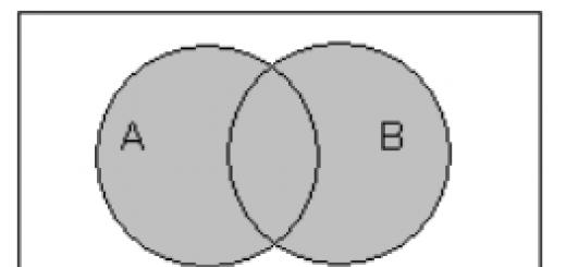
Pavlov proved that cortical end of analyzer This is not a strictly defined area. It has a core and scattered elements. Core- a place of concentration of nerve cells, where the highest analysis, synthesis and integration take place. On its periphery, in scattered elements, simple analysis and synthesis are carried out. The areas of scattered elements of adjacent analyzers overlap each other (Fig.).
According to Pavlov, the work of the second signaling system is inextricably linked with the functions of all analyzers, therefore it is impossible to imagine the localization of the complex functions of the second signaling system in limited cortical fields. Pavlov laid the foundations for the doctrine of the dynamic localization of functions in the cortex. The concept of dynamic localization of functions in the cortex suggests the possibility of using the same cortical structures in various combinations to serve various complex cortical functions. Thus, associative paths unite analyzers, contributing to the higher synthetic activity of the cerebral cortex. Today, scientists know that irritation is transformed into excitation, which is transmitted to the cortical end of the analyzer. Another thing is not clear - where and how does excitement transform into sensation? What structures are responsible for this? So, when the visual field is irritated in the region of the spur groove, "simple" hallucinations appear in the form of light or color spots, sparks, shadows. Irritation of the outer surface of the occipital lobe gives "complex" hallucinations in the form of figures, moving objects.
In the motor zone of the cortex, cells were found that give a discharge of impulses to visual, auditory, and skin stimuli, and in the visual zone of the cortex, neurons were found that respond with electrical discharges to tactile, auditory, vestibular, and olfactory stimuli. In addition, neurons have been found that respond not only to "their own" stimulus, as they say now, the stimulus of their modality, their quality, but also to one or two others. They are called polysensory neurons.
This section of the anatomy of the NS is divided into the following subcategories
Ideas about the localization of functions in the cerebral cortex are of great practical importance for solving problems of the topic of lesions in the cerebral hemispheres. However, to this day, much in this section remains controversial and not fully resolved. The doctrine of the localization of functions in the cortex has a rather long history - from the denial of the localization of functions in it to the distribution in the cortex in strictly limited territories of all the functions of human activity, up to the highest qualities of the latter (memory, will, etc.), and, finally , until returning to the "equipotentiality" of the cortex, that is, again, in essence, to the denial of the localization of functions (recently abroad).
Ideas about the equivalence (equipotentiality) of various cortical fields come into conflict with the huge factual material accumulated by morphologists, physiologists and clinicians. Everyday clinical experience shows that there are certain unshakable regular dependences of functional disorders on the location of the pathological focus. Based on these basic provisions, the clinician solves the problems of topical diagnosis. However, this is the case as long as we operate with disorders related to relatively simple functions: movements, sensitivity, etc. parts of the nervous system and the periphery. The functions of the cortex are more complex, phylogenetically younger, and cannot be narrowly localized; very extensive areas of the cortex, and even the entire cortex as a whole, are involved in the implementation of complex functions. That is why the solution of problems of the topic of lesions based on speech disorders, apraxia, agnosia, and, moreover, mental disorders, as clinical experience shows, is more difficult and sometimes inaccurate.
At the same time, within the cerebral cortex there are areas whose damage causes one or another character, one or another degree, for example, speech disorders, disorders of gnosia and praxia, the topodiagnostic value of which is also significant. From this, however, it does not follow that there are special, narrowly localized centers that "manage" these most complex forms of human activity. It is necessary to clearly distinguish between the localization of functions and the localization of symptoms.
The foundations of a new and progressive theory of the localization of functions in the brain were created by I.P. Pavlov.
Instead of the concept of the cerebral cortex as, to a certain extent, an isolated superstructure over other floors of the nervous system with narrowly localized areas connected along the surface (associative) and with the periphery (projection) areas, I.P. Pavlov created the doctrine of the functional unity of neurons belonging to various parts of the nervous system - from receptors on the periphery to the cerebral cortex - the doctrine of analyzers. What we call the center is the highest, cortical, section of the analyzer. Each analyzer is associated with certain areas of the cerebral cortex (Fig. 64).
I.P. Pavlov makes significant adjustments to the previous ideas about the limited territories of the cortical centers, to the doctrine of the narrow localization of functions. Here is what he says about the projection of receptors into the cerebral cortex.
“Each peripheral receptor apparatus has a central, special, isolated territory in the cortex, as its terminal station, which represents its exact projection. Here, thanks to a special design, there can be a denser placement of cells, more numerous cell connections and the absence of cells of other functions, the most complex irritations occur, form (higher synthesis) and their exact differentiation (higher analysis) takes place. But these receptor elements also spread further over a very long distance, perhaps throughout the cortex.” This conclusion, based on extensive experimental and physiological studies, is in full agreement with the latest morphological data on the impossibility of precise differentiation of cortical cyto-architectonic fields.
Consequently, the functions of the analyzers (or, in other words, the operation of the first signaling system) cannot be associated only with the cortical projection zones (the nuclei of the analyzers). Moreover, it is impossible to narrowly localize the most complex, purely human functions - the functions of the second signaling system.
I.P. Pavlov defines the functions of human signaling systems as follows. “I imagine the totality of higher nervous activity in this way. In higher animals, up to and including humans, the first instance for the complex relationships of the organism with the environment is the subcortex closest to the hemispheres with its most complex unconditioned reflexes (our terminology), instincts, drives, affects, emotions (diverse, usual terminology). These reflexes are caused by relatively few unconditional external agents. Hence - a limited orientation in the environment and at the same time a weak adaptation.
The second instance is the large hemispheres ... Here, with the help of a conditional connection (association), a new principle of activity arises: the signaling of a few, unconditional external agents by an innumerable mass of other agents, at the same time constantly analyzed and synthesized, making it possible to have a very large orientation in the same environment and by the same much more fitting. This constitutes the only signaling system in the animal body and the first in man.
In a person, another signaling system is added, signaling the first system with speech, its basis or basal component - kinesthetic stimuli of the speech organs. This introduces a new principle of nervous activity - the abstraction and together the generalization of countless signals of the previous system, in turn, again with the analysis and synthesis of these first generalized signals - the principle that determines an unlimited orientation in the surrounding world and creates the highest adaptation of man - science, as in the form of a universal empiricism, as well as in its specialized form.
The work of the second signal system is inextricably linked with the functions of all analyzers, therefore it is impossible to imagine the localization of the complex functions of the second signal system in any limited cortical fields.
The significance of the legacy left to us by the great physiologist for the correct development of the doctrine of the localization of functions in the cerebral cortex is exceptionally great. I.P. Pavlov laid the foundations for a new theory of dynamic localization of functions in the cortex. The concept of dynamic localization implies the possibility of using the same cortical structures in various combinations to serve various complex cortical functions.
Keeping a number of definitions and interpretations that have become established in the clinic, we will try to make some adjustments to our presentation in the light of the teachings of I.P. Pavlov about the nervous system and its pathology.
So, first of all, we need to consider the question of the so-called projection and association centers. The usual idea of motor, sensory and other projection centers (anterior and posterior central gyrus, visual, auditory centers, etc.) is associated with the concept of a rather limited localization of a particular function in a given area of the cortex, and this center is directly connected with the underlying nerve devices , and subsequently with the periphery, with its conductors (hence the definition - "projective"). An example of such a center and its conductor is, for example, the anterior central gyrus and the pyramidal path; fissura calcarina and radiatio optica, etc. Projection centers are connected by associative paths with other centers, with the surface of the cortex. These wide and powerful associative pathways determine the possibility of the combined activity of various cortical areas, the establishment of new connections, and the formation, therefore, of conditioned reflexes.
"Association centers", in contrast to projection centers, do not have a direct connection with the underlying parts of the nervous system and the periphery; they are connected only with other areas of the cortex, including the "projection centers". An example of an "association center" is the so-called "center of stereognosy" in the parietal lobe, located posterior to the posterior central gyrus (Fig. 65). Individual stimuli that occur when an object is felt by hand enter the posterior central gyrus through the thalamo-cortical pathways: tactile, shapes and sizes (joint-muscular feeling), weight, temperature, etc. All these sensations are transmitted through association fibers from the posterior central gyrus to the "stereognostic center", where they are combined and create a common sensory image of the object. The connections of the “stereognostic center” with other areas of the cortex make it possible to identify, compare this image with the idea already in memory of this object, its properties, purpose, etc. (i.e., analysis and synthesis of perception is carried out). This "center", therefore, has no direct connection with the underlying parts of the nervous system and is connected by association fibers with a number of other fields of the cerebral cortex.
The division of centers into projection and association seems to us incorrect. The large hemispheres are a set of analyzers for analyzing, on the one hand, the external world and, on the other, internal processes. The receptive centers of the cortex appear to be very complicated and territorially extremely widespread. The upper layers of the cerebral cortex, in fact, are entirely occupied by the perceiving centers or, in the terminology of I.P. Pavlov, "brain ends of analyzers".
From all the lobes, from the lower layers of the cortex, there are already efferent conductors connecting the cortical ends of the analyzers with the executive organs through the subcortical, stem and spinal apparatuses. An example of such an efferent conductor is the pyramidal pathway - this intercalary neuron between the kinesthetic (motor) analyzer and the peripheral motor neuron.
How, then, from this point of view, to reconcile the position about the presence of motor projection centers (in the anterior central gyrus, the center of eye rotation, etc.), when they are turned off, a person experiences paralysis, and when irritated, convulsions with a completely clear somatotopic distribution and correspondence? Here we are talking only about the defeat of the motor projection area for the pyramidal pathways, and not the "projection motor centers".
There is no doubt that "voluntary" movements are conditioned motor reflexes, that is, movements that have developed, "trodden" in the process of individual life experience: but in the development, organization and already established activity of skeletal muscles, everything depends on the afferent device - skin and motor analyzer (clinically - skin and joint-muscular sensitivity, more broadly - kinesthetic sense), without which fine and precise coordination of a motor act is impossible.
Rice. 64. Cortical departments of analyzers (scheme).
a - outer surface; b - inner surface. Red - skin analyzer; yellow - auditory analyzer: blue - visual analyzer; green - olfactory analyzer; dotted line - motor analyzer.
The motor analyzer (whose task is the analysis and synthesis of “voluntary” movements) does not at all correspond to the ideas of cortical motor “projection” centers with certain boundaries of the latter and a clear somatotopic distribution. The motor analyzer, like all analyzers, is associated with very wide areas of the cortex, and the motor function (in relation to "voluntary" movements) is extremely complex (if we take into account not only the determinism of movements and behavior in general, not only the complexity of action complexes, but also afferent kinesthetic systems , and orientation in relation to the environment and parts of one's own body in space, etc.).
What is the idea of "projection centers"? It was argued that the latter represent a kind of input or output "starting gate" for impulses coming into or out of the cortex. And if we accept that “motor projection cortical centers” are only such “gates” (for the broad concept of a motor analyzer is necessarily associated with the function of analysis and synthesis), then it should be considered that within the anterior central gyrus (and in territories similar to it), and then only in certain of its layers, there is a motor projection area or zone.
How then to imagine the rest of the "projection" centers (skin sensitivity, vision, hearing, taste, smell) associated with other (non-kinesthetic) afferent systems? We think that there is no fundamental difference here: in fact, impulses from the periphery, which occurs within many layers and wide areas.
Consequently, in each analyzer (its cortical section), including the motor one, there is an area or zone that “projects” onto the periphery (motor area) or into which the periphery is “projected” (sensitive areas, including kinesthetic receptors for the motor analyzer ).
It is possible that the "projective core of the analyzer" can be identified with the concept of a motor or sensitive projection zone. Maximum violations, wrote I.I. Pavlov, analysis and synthesis occurs when just such a “projective nucleus” is damaged; if. If we take for the real maximum "breakdown" of the analyzer the maximum impairment of function, which is objectively absolutely correct, then the greatest manifestation of damage to the motor analyzer is central paralysis, and that of the sensory analyzer is anesthesia. From this point of view, it would be correct to identify the concept of “analyzer core” with the concept of “analyzer projection area”.
Rice. 65. Loss of functions observed in the defeat of various parts of the cerebral cortex (outer surface).
2 - visual disturbances (hemianopsia); 3 - sensitivity disorders; 4 - central paralysis or paresis; 5 - agraphia; 6 - cortical paralysis of gaze and turning of the head in the opposite direction; 7 - motor aphasia; 8 - hearing disorders (with unilateral lesions are not observed); 9 - amnestic aphasia; 10 - alexia; 11 - visual agnosia (with bilateral lesions); 12 - astereognosia; 13 - apraxia; 14 - sensory aphasia.
Based on the foregoing, we consider it correct to replace the concept of a projection center with the concept of a projection area in the analyzer zone. Then the division of cortical "centers" into projection and association ones is unreasonable: there are analyzers (their cortical sections) and within their limits - projection areas.
Lecture 12 Projection cortical zones: primary and secondary. Motor (motor) zones of the cerebral cortex. Tertiary cortical zones.
 Loss of functions observed when various parts of the cortex (inner surface) are affected. 1 - olfactory disorders (with unilateral lesions are not observed); 2 - visual disturbances (hemianopsia); 3 - sensitivity disorders; 4 - central paralysis or paresis. The data of experimental studies on the destruction or removal of certain areas of the cortex and clinical observations indicate the confinement of functions to the activity of certain areas of the cortex. A section of the cerebral cortex that has some specific function is called the cortical zone. There are projection, associative cortical zones and motor (motor).
 The projection cortical zone is the cortical representation of the analyzer. The neurons of the projection zones receive signals of one modality (visual, auditory, etc.). Distinguish: - primary projection zones; - secondary projection zones providing an integrative function of perception. In the zone of one or another analyzer, tertiary fields, or associative zones, are also distinguished.
 The primary projection fields of the cortex receive information mediated through the smallest number of switches in the subcortex (in the thalamus, diencephalon). On these fields, the surface of peripheral receptors is, as it were, projected. Nerve fibers enter the cerebral cortex mainly from the thalamus (these are afferent inputs).
 The projection zones of the analyzer systems occupy the outer surface of the cortex of the posterior parts of the brain. This includes the visual (occipital), auditory (temporal) and general sensory (parietal) areas of the cortex. The cortical department also includes the representation of taste, olfactory, visceral sensitivity
 Primary sensory areas (Brodmann fields): visual - 17, auditory - 41 and somatosensory - 1, 2, 3 (collectively they are called sensory cortex), motor (4) and premotor (6) cortex
 Primary sensory areas (Brodmann fields): visual - 17, auditory - 41 and somatosensory - 1, 2, 3 (collectively they are called sensory cortex), motor (4) and premotor (6) cortex Each field of the cerebral cortex is characterized by a special composition neurons, their location and connections between them. The fields of the sensory cortex, in which the primary processing of information from sensory organs takes place, differ sharply from the primary motor cortex, which is responsible for the formation of commands for voluntary muscle movements.
 The motor cortex is dominated by neurons resembling pyramids in shape, and the sensory cortex is represented mainly by neurons whose body shape resembles grains, or granules, which is why they are called granular. The structure of the cerebral cortex I. molecular II. external granular III. external pyramidal IV. internal granular V. ganglionic (giant pyramids) VI. polymorphic
 Neurons of the primary projection zones of the cortex, which mainly have the highest specificity. For example, the neurons of the visual areas selectively respond to shades of color, the direction of movement, the nature of the lines, etc. However, in the primary zones of individual areas of the cortex there are also multimodal neurons that respond to several types of stimuli and neurons whose reaction reflects the impact of nonspecific ( limbicoreticular) systems.
 The projection afferent fibers terminate in the primary fields. So, fields 1 and 3, occupying the medial and lateral surface of the posterior central gyrus, are the primary projection fields of the skin sensitivity of the body surface.
 The functional organization of the projection zones in the cortex is based on the principle of topical localization. Perceptive elements located next to each other on the periphery (for example, skin areas) are projected onto the cortical surface also next to each other.
 In the medial part, the lower limbs are represented, and the projections of the receptor fields of the skin surface of the head are located the lowest on the lateral part of the gyrus. At the same time, areas of the body surface richly supplied with receptors (fingers, lips, tongue) are projected onto a larger area of the cortex than areas with fewer receptors (thigh, back, shoulder).

 Fields 17-19, located in the occipital lobe, are the visual center of the cortex, the 17th field, which occupies the occipital pole itself, is primary. The 18th and 19th fields adjacent to it perform the function of secondary fields and receive inputs from the 17th field.
 The auditory projection fields are located in the temporal lobes (41, 42). Next to them, on the border of the temporal, occipital and parietal lobes, are the 37th, 39th and 40th, which are characteristic only of the human cerebral cortex. For most people, in these fields of the left hemisphere, the speech center is located, which is responsible for the perception of oral and written speech.

 Secondary projection fields that receive information from the primary ones are located next to them. The neurons of these fields are characterized by the perception of complex signs of stimuli, but at the same time, the specificity corresponding to the neurons of the primary zones is preserved. The complication of the detector properties of neurons in the secondary zones can occur by convergence of neurons in the primary zones on them. In the secondary zones (the 18th and 19th Brodmann fields), detectors of more complex contour elements appear: edges of a limited length of lines, angles with different orientations, etc.
 Motor (motor) zones of the cerebral cortex are areas of the motor cortex, the neurons of which cause a motor act. The motor areas of the cortex are located in the precentral gyrus of the frontal lobe (in front of the projection zones of skin sensitivity). This part of the cortex is occupied by fields 4 and 6. From the V layer of these fields, the pyramidal path originates, ending at the motor neurons of the spinal cord.
 Premotor zone (field 6) The premotor zone of the cortex is located in front of the motor zone, it is responsible for muscle tone and performing coordinated movements of the head and trunk. The main efferent outputs from the cortex are the axons of the layer V pyramids. These are efferent, motor neurons involved in the regulation of motor functions.


 Tertiary or interanalyzer zones (associative) Prefrontal zone (fields 9, 10, 45, 46, 47, 11), parietotemporal (fields 39, 40) Afferent and efferent projection zones of the cortex occupy a relatively small area. Most of the surface of the cortex is occupied by tertiary or interanalyzer zones, called associative. They receive polymodal inputs from sensory areas of the cortex and thalamic associative nuclei and have outputs to the motor areas of the cortex. Associative zones provide integration of sensory inputs and play an essential role in mental activity (learning, thinking).
 Functions of different zones of the new cortex: 5 3 7 6 4 1 2 Memory, needs Behavior triggering 1. Occipital lobe - visual cortex. 2. Temporal lobe - auditory cortex. 3. Anterior part of the parietal lobe - pain, skin and muscle sensitivity. 4. Inside the lateral sulcus (insular lobe) - vestibular sensitivity and taste. 5. The back of the frontal lobe is the motor cortex. 6. The back of the parietal and temporal lobes - the associative parietal cortex: combines signal flows from different sensory systems, speech centers, thought centers. 7. The anterior part of the frontal lobe - the associative frontal cortex: taking into account sensory signals, signals from the centers of needs, memory and thinking, makes decisions to launch behavioral programs ("center of will and initiative").
 Separate large associative areas are located next to the corresponding sensory areas. Some associative zones perform only a limited specialized function and are associated with other associative centers capable of subjecting information to further processing. For example, the audio association area analyzes sounds into categories and then relays signals to more specialized areas, such as the speech association area, where the meaning of the words heard is perceived.
 The associative fields of the parietal lobe combine information coming from the somatosensory cortex (from the skin, muscles, tendons and joints regarding body position and movement) with visual and auditory information coming from the visual and auditory cortex of the occipital and temporal lobes. This combined information helps to have an accurate picture of one's own body while moving around in the environment.
 Wernicke's area and Broca's area are two areas of the brain involved in the process of reproducing and understanding information related to speech. Both areas are located along the Sylvian sulcus (lateral sulcus of the cerebral hemispheres). Aphasia is a complete or partial loss of speech due to local lesions of the brain.


Subsequently, the efforts of physiologists turned out to be aimed at finding "critical" areas of the brain, the destruction of which led to a violation of the reflex activity of one or another organ. Gradually, an idea was formed about the rigid anatomical localization of "reflex arcs", and, accordingly, the reflex itself began to be thought of as a mechanism for the operation of only the lower parts of the brain (spinal centers).
At the same time, the question of the localization of functions in the higher parts of the brain was being developed. Ideas about the localization of elements of mental activity in the brain arose long ago. In almost every era, one or more
Other hypotheses of representation in the brain of higher mental functions and consciousness in general.
Austrian physician and anatomist Franz Joseph Gall(1758-1828) compiled a detailed description of the anatomy and physiology of the human nervous system, provided with an excellent atlas.
: A whole generation of researchers have been based on these data. Gall's anatomical discoveries include the following: identification of the main differences between the gray and white matter of the brain; determination of the origin of nerves in the gray matter; definitive proof of decussation of the pyramidal tracts and optic nerves; establishment of differences between "convergent" (according to modern terminology "associative") and "divergent" ("projective") fibers (1808); the first clear description of the commissures of the brain; proof of the beginning of the cranial nerves in the medulla oblongata (1808), etc. Gall was one of the first to give a decisive role to the cerebral cortex in the functional activity of the brain. Thus, he believed that the folding of the cerebral surface is an excellent solution by nature and evolution of an important task: to ensure the maximum increase in the surface area of the brain while maintaining its volume more or less constant. Gall introduced the term "arc", familiar to every physiologist, and described its clear division into three parts.
However, Gall's name is mostly known in connection with his rather dubious (and sometimes scandalous!) doctrine of the localization of higher mental functions in the brain. Attaching great importance to the correspondence of function and structure, Gall as early as 1790 made an application for the introduction of a new science into the arsenal of knowledge - phrenology(from the Greek phren - soul, mind, heart), which also received a different name - psychomorphology, or narrow localizationism. As a doctor, Gall observed patients with various disorders of brain activity and noticed that the specificity of the disease largely depends on which part of the brain substance is damaged. This led him to the idea that each mental function corresponds to a specific part of the brain. Seeing the infinite variety of characters and individual mental qualities of people, Gall suggested that the strengthening (or greater predominance) of any character trait or mental function in a person’s behavior also entails the predominant development of a certain area of the cerebral cortex where this function is represented. Thus, the thesis was put forward: the function makes the structure. As a result of the growth of this hypertrophied area of the cortex ("brain cones"), pressure on the bones of the skull increases, which, in turn, causes the appearance of an external cranial tubercle above the corresponding area of \u200b\u200bthe brain. In case of underdevelopment of the function, vice versa.
On the surface of the skull there will be a noticeable depression ("fossa"). Using the method of "cranioscopy" created by Gall - the study of the relief of the skull with the help of palpation - and detailed "topographic" maps of the surface of the brain, which indicated the places of localization of all abilities (considered innate), Gall and his followers made a diagnosis, i.e. made a conclusion about character and inclinations of a person, about his mental and moral qualities. Were 2 allocated? areas of the brain where certain abilities of the individual are localized (moreover, 19 of them were recognized as common to humans and animals, and 8 as purely human). In addition to the "bumps" responsible for the implementation of physiological functions, there were those that testified to visual and auditory memory, orientation in space, a sense of time, the instinct of procreation; such personality traits. as courage, ambition, piety, wit, secrecy, amorousness, caution, self-esteem, refinement, hope, curiosity, malleability of education, pride, independence, diligence, aggressiveness, fidelity, love of life, love of animals.
Gall's erroneous and pseudoscientific ideas (which, however, were extremely popular in their time) contained a rational grain: the recognition of the closest connection between the manifestations of mental functions and the activity of the cerebral cortex. The problem of finding differentiated "think tanks" and drawing attention to the functions of the brain was put on the agenda. Gall can truly be considered the founder of "brain localization". Of course, for the further progress of psychophysiology, the formulation of such a problem was more promising than the ancient search for the location of the "common sensory area".
The solution of the question of the localization of functions in the cerebral cortex was facilitated by data accumulating in clinical practice and in experiments on animals. German physician, anatomist and physicist Julius Robert Mayer(1814-1878), who practiced for a long time in Parisian clinics, and also served as a ship's doctor, observed in patients with craniocerebral injuries the dependence of a violation (or complete loss) of a particular function on damage to a certain part of the brain. This allowed him to suggest that memory is localized in the cerebral cortex (it should be noted that T. Willis came to a similar conclusion back in the 17th century), imagination and judgments in the white matter of the brain, apperception and will in the basal ganglia. A kind of "integral organ" of behavior and the psyche is, according to Mayer, the corpus callosum and the cerebellum.
Over time, the clinical study of the consequences of brain damage was supplemented by laboratory studies. artificial extirpation method(from Latin ex (s) tirpatio - removal with a root), which allows you to partially or completely destroy (remove) parts of the brain of animals to determine their functional role in brain activity. At the beginning of the XIX century. carried out mainly acute experiments on animals (frogs, birds), later, with the development of asepsis methods, chronic experiments began to be carried out, which made it possible to observe the behavior of animals for a more or less long time after the operation. Removal of various parts of the brain (including the cerebral cortex) in mammals (cats, dogs, monkeys) made it possible to elucidate the structural and functional basis of complex behavioral reactions.
It turned out that the deprivation of animals of the higher parts of the brain (birds - the forebrain, mammals - the cerebral cortex) in general did not cause a violation of the main functions: respiration, digestion, excretion, blood circulation, metabolism and energy. Animals retained the ability to move, to respond to certain external influences. Consequently, the regulation of these physiological manifestations of vital activity occurs at the underlying (compared to the cerebral cortex) levels of the brain. However, when the higher parts of the brain were removed, profound changes in the behavior of animals occurred: they became practically blind and deaf, “stupid”; they lost previously acquired skills and could not develop new ones, could not adequately navigate in the environment, did not distinguish and could not differentiate objects in the surrounding space. In a word, animals became "living automata" with monotonous and rather primitive ways of responding.
In experiments with partial removal of areas of the cerebral cortex, it was found that the brain is functionally heterogeneous and the destruction of one area or another leads to a violation of a certain physiological function. So, it turned out that the occipital areas of the cortex are associated with visual function, the temporal - with auditory, the region of the sigmoid gyrus - with motor function, as well as with skin and muscle sensitivity. Moreover, this differentiation of functions in individual regions of the higher parts of the brain is being improved in the course of the evolutionary development of animals.
The strategy of scientific research in the study of brain functions led to the fact that, in addition to the method of extirpation, scientists began to use the method of artificial stimulation of certain areas of the brain using electrical stimulation, which also made it possible to evaluate the functional role of the most important parts of the brain. The data obtained using these methods of laboratory research, as well as the results of clinical observations, outlined one of the main directions of psychophysiology in the 19th century. - determination of the localization of nerve centers responsible for higher mental functions and behavior of the organism as a whole. So. in 1861, the French scientist, anthropologist and surgeon Paul Broca (1824-1880), on the basis of clinical facts, strongly opposed the physiological equivalence of the cerebral cortex. During the autopsy of the corpses of patients suffering from a speech disorder in the form of motor aphasia (the patients understood someone else's speech, but they themselves could not speak), he found changes in the posterior part of the lower (third) frontal gyrus of the left hemisphere or in the white matter under this area of the cortex. Thus, as a result of these observations, Broca established the position of the motor (motor) center of speech, later named after him. In 1874, the German psychiatrist and neurologist K? Wernicke (1848-1905) described the sensory center of speech (today bearing his name) in the posterior third of the first temporal gyrus of the left hemisphere. The defeat of this center leads to the loss of the ability to understand human speech (sensory aphasia). Even earlier, in 1863, using the method of electrical stimulation of certain areas of the cortex (the precentral gyrus, the precentral region, the anterior part of the pericentral lobule, the posterior parts of the upper and middle frontal gyri), the German researchers Gustav Fritsch and Eduard Gitzig established motor centers (motor cortical fields), the irritation of which caused certain contractions of the skeletal muscles, "and the destruction led to profound disorders of motor behavior. In 4874, the Kiev anatomist and physician Vladimir Alekseevich Betz (1834-1894) discovered efferent nerve cells of the motor centers - giant pyramidal cells of the V layer cortex, named after him Betz cells German researcher Hermann Munch (student of I. Müller and E. Dubois-Reymond) discovered not only the motor cortical fields, using the method of extirpation, he found the centers of sensory perceptions He managed to show that the center of vision is located in the posterior lobe of the brain, c the center of hearing is in the temporal lobe. Removal of the occipital lobe of the brain led to the loss of the animals' ability to see (with complete preservation of the visual apparatus). Already in
early 20th century eminent Austrian neurologist Konstantin Economo(1876-1931) the centers of swallowing and chewing were established in the so-called black matter of the brain (1902), the centers that control sleep - in the midbrain (1917). Running a little ahead, we say that Economo gave an excellent description of the structure of the cerebral cortex an adult and in 1925 refined the cytoarchitectonic map of the cortical fields of the brain, putting 109 fields on it.
However, it should be noted that in the XIX century. serious arguments were put forward against the position of narrow localizationists, according to whose views motor and sensory functions are confined to different areas of the cerebral cortex. Thus, a theory of the equivalence of sections of the cortex arose, asserting the idea of the equal importance of cortical formations for the implementation of any activity of the body, - equipotentialism. In this regard, the phrenological views of Gall, one of the most vehement supporters of localizationism, were criticized by the French physiologist Marie Jean Pierre Flourance(1794-1867). Back in 1822, he pointed out the presence of a respiratory center in the medulla oblongata (which he called the "vital knot"); linked coordination of movements with the activity of the cerebellum, vision - with the quadrigemina; I saw the main function of the spinal cord in conducting excitation along the nerves. However, despite such seemingly localizationist views, Flurence believed that the basic mental processes (including intellect and will) underlying the purposeful behavior of a person are carried out as a result of the activity of the brain as a holistic formation and therefore a holistic behavioral function. cannot be confined to any particular anatomical entity. Flourance spent most of his experiments on pigeons and chickens, removing parts of their brains and observing changes in the behavior of birds. Usually, after some time after the operation, the behavior of birds was restored regardless of which areas of the brain were damaged, so Flurance concluded that the degree of violation of various forms of behavior is determined primarily by how much brain tissue was removed during the operation. Having improved the technique of operations, he was the first to be able to completely remove the hemispheres of the forebrain from animals and save their life for further observations.
Based on the experiments, Flurance came to the conclusion that the forebrain hemispheres play a decisive role in the implementation of a behavioral act. Their complete removal leads to the loss of all "intelligent" functions. Moreover, especially severe behavioral disorders were observed in chickens after the destruction of the gray matter of the surface of the cerebral hemispheres - the so-called corticoid plate, an analogue of the cerebral cortex of mammals. Flourance suggested that this area of the brain is the seat of the soul, or "controlling spirit", and therefore acts as a whole, having a homogeneous and equivalent mass (similar, for example, to the tissue structure of the liver). Despite the somewhat fantastic ideas of the equipotentialists, one should note the progressive element in their views. First, complex psychophysiological functions were recognized as the result of the combined activity of brain formations. Secondly, the idea of a high dynamic plasticity of the brain, expressed in the interchangeability of its parts, was put forward.
- Gall managed to quite accurately determine the "center of speech", but "officially" it was discovered by the French researcher Paul Broca (1861).
- In 1842 Mayer, working on the definition of the mechanical equivalent of heat, came to a generalized law of conservation of energy.
- Unlike his predecessors, who endowed the nerve with the ability to feel (ie, recognizing a certain mental quality behind it), Hall considered the nerve ending (in the sense organ) to be an "apsychic" formation.
This question is extremely important theoretically and especially practically. Hippocrates already knew that brain injuries lead to paralysis and convulsions on the opposite side of the body, and sometimes are accompanied by loss of speech.
In 1861, the French anatomist and surgeon Broca, on autopsy of the corpses of several patients suffering from a speech disorder in the form of motor aphasia, discovered profound changes in the pars opercularis of the third frontal gyrus of the left hemisphere or in the white matter under this area of the cortex. Based on his observations, Broca established in the cerebral cortex the motor center of speech, later named after him.
The English neuropathologist Jackson (1864) spoke in favor of the functional specialization of individual sections of the hemispheres on the basis of clinical data. Somewhat later (1870), the German researchers Fritsch and Gitzig proved the existence of special areas in the dog's cerebral cortex, the stimulation of which by a weak electric current is accompanied by a contraction of individual muscle groups. This discovery caused a large number of experiments, basically confirming the existence of certain motor and sensory areas in the cerebral cortex of higher animals and humans.
On the issue of localization (representation) of function in the cerebral cortex, two diametrically opposed points of view competed with each other: localizationists and antilocalizationists (equipotentialists).
Localizationists were supporters of narrow localization of various functions, both simple and complex.
The anti-localizationists took a completely different view. They denied any localization of functions in the brain. The whole bark for them was equivalent and homogeneous. All its structures, they believed, have the same possibilities for performing various functions (equipotential).
The problem of localization can be correctly resolved only with a dialectical approach to it, which takes into account both the integral activity of the entire brain and the different physiological significance of its individual parts. It was in this way that IP Pavlov approached the problem of localization. In favor of the localization of functions in the cortex, numerous experiments by IP Pavlov and his colleagues with the extirpation of certain areas of the brain convincingly speak. Resection of the occipital lobes of the cerebral hemispheres (centers of vision) in a dog causes enormous damage to the conditioned reflexes developed in it to visual signals and leaves intact all conditioned reflexes to sound, tactile, olfactory and other stimuli. On the contrary, resection of the temporal lobes (hearing centers) leads to the disappearance of conditioned reflexes to sound signals and does not affect the reflexes associated with optical signals, etc. Against equipotentialism, in favor of the representation of the function in certain areas of the cerebral hemispheres, the latest data from electroencephalography also speak . Irritation of a certain area of the body leads to the appearance of reactive (evoked) potentials in the cortex in the "center" of this area.
IP Pavlov was a staunch supporter of localization of functions in the cerebral cortex, but only relative and dynamic localization. The relativity of localization is manifested in the fact that each section of the cerebral cortex, being the carrier of a certain special function, the "center" of this function, responsible for it, participates in many other functions of the cortex, but not as the main link, not in the role of the "center ”, but on a par with many other areas.
The functional plasticity of the cortex, its ability to restore the lost function by establishing new combinations speak not only of the relativity of the localization of functions, but also of its dynamism.
The basis of any more or less complex function is the coordinated activity of many areas of the cerebral cortex, but each of these areas participates in this function in its own way.
The basis of modern ideas about the "systemic localization of functions" is the teaching of I. P. Pavlov about the dynamic stereotype. Thus, the higher mental functions (speech, writing, reading, counting, gnosis, praxis) have a complex organization. They are never carried out by some isolated centers, but are always processes "placed in a complex system of zones of the cerebral cortex" (AR Luria, 1969). These "functional systems" are mobile; in other words, the system of means by which this or that task can be solved changes, which, of course, does not reduce the importance for them of the well-studied “fixed” cortical areas of Broca, Wernicke, and others.
The centers of the cerebral cortex of a person are divided into symmetrical, presented in both hemispheres, and asymmetric, present in only one hemisphere. The latter include the centers of speech and functions associated with the act of speech (writing, reading, etc.), which exist only in one hemisphere: in the left - in right-handers, in the right - in left-handers.
Modern ideas about the structural and functional organization of the cerebral cortex come from the classical Pavlovian concept of analyzers, refined and supplemented by subsequent studies. There are three types of cortical fields (G. I. Polyakov, 1969). The primary fields (analyzer cores) correspond to the architectonic zones of the cortex, where sensory pathways end (projection zones). Secondary fields (peripheral sections of the analyzer nuclei) are located around the primary fields. These zones are connected with receptors indirectly, in them a more detailed processing of incoming signals takes place. Tertiary, or associative, fields are located in zones of mutual overlap of the cortical systems of analyzers and occupy more than half of the entire surface of the cortex in humans. In these zones, inter-analyzer connections are established that provide a generalized form of a generalized action (V. M. Smirnov, 1972). The defeat of these zones is accompanied by violations of gnosis, praxis, speech, purposeful behavior.