Application of radioactive isotopes in industry, science and agriculture (according to press data)// Atomic Energy. Volume 2, issue. 1. - 1957. - pp. 85-88.
APPLICATION OF RADIOACTIVE ISOTOPES IN INDUSTRY, SCIENCE AND AGRICULTURE
(According to PRINT DATA)
Ferrous metallurgy
At the Stalin Metallurgical Plant, using radiocobalt contained in a small container, the welds of the blast furnace casing were scanned directly during its installation, i.e., in conditions where X-ray photography was completely impossible. Using such containers, flaw detectors detect internal defects in the walls of steam boilers, gas pipelines, etc.
The duration of smelting in open hearth furnaces and the purification of steel from sulfur and phosphorus at the early stage of smelting largely depend on the order of charging and the amount of solid materials. Using radioisotopes, a number of plants “tag” charge materials—lime or ore—and by measuring the radioactivity of metal or slag samples, they determine the rate of active slag formation. In this way, the dependence of the dissolution rate is revealed
limestone and ore from technological factors. The melting rate of scrap is determined in the same way. To do this, the metal part of the charge is “marked”. Such research is currently being carried out at the Azovstal, Stalinsky, Makeevsky and Magnitogorsk and Kuznetsk plants.
After producing steel in an open-hearth furnace, it is not always possible to assess the condition of the welding of the hearth by means of a routine inspection. Meanwhile, an undetected defect can lead to a serious accident in subsequent heats. Radioisotopes are used to prevent this. At the Azov Steel plant, radiophosphorus in magnesite ampoules was placed in various places in the hearth. The appearance of radioactivity in slag samples taken along the melting process indicated the destruction of the welding.
In the field of analytical chemistry in relation to metallurgical problems, two directions of research using radioactive isotopes have emerged. The first is the testing and refinement of conventional chemical analysis methods, such as phosphorus. In this case, when dissolving a sample of steel or ore, a small amount of radiophosphorus is added. During the analysis, at its various stages, the intensity of radioactive radiation of the solution samples is determined. In the absence of losses, the sum of the radioactivity of solutions taken at all stages of analysis should be equal to the radioactivity of the original solution.
The second direction is the development of methods for express analysis of steel and slag for any one element, the most important for this technology, by introducing radioactive isotopes directly into steel-smelting furnaces. Thus, for the technology of processing cast iron with a high phosphorus content, the Azovstal plant has developed a method for the rapid determination of phosphorus pentoxide in slags. It has been established that with a consumption of 0.04-0.05 microcuries of radioactive phosphorus isotope per ton of metal, sufficient analysis accuracy is achieved with a significantly shorter duration compared to chemical analysis. This method allows you to control the slag melting regime and more accurately sort the slag as fertilizer. A similar method has been developed for determining phosphorus in metal during the blowing of pig iron in a converter. It can be successfully used in developing new metallurgical processing technology and in research where the speed of determining the phosphorus content in metal or slag affects the course of the process. (Industrial Economic Newspaper, September 9, 1956)
In the struggle for technical progress, Ukrainian metallurgists are increasingly using labeled atoms of radioactive elements. With the help of radioactive cobalt, without stopping the blast furnace, the condition of its walls, lined with refractory bricks, is determined.
Using radioactive isotopes, open-hearth workers establish the rate at which crystallization of steel ingots occurs, determine the phosphorus content in open-hearth slag many times faster than before, observe the process of slag formation, and record the moment of completion of the process of melting iron ore and limestone during smelting.
Using labeled atoms, metallurgists determine the silica content in ore several times faster than by chemical methods. Radioactive isotopes are widely used to determine the thickness and nature of the occurrence of iron ore layers and to control a number of production processes at crushing, screening and processing plants. (Pravda Ukrainy, Aug. 4, 1956)
#At the Zaporozhye plant “Dneprospetsstal” a laboratory of physical research methods, equipped with the latest equipment, came into operation. This laboratory conducts research into the process of smelting electric steel using radioactive isotopes. In particular, the cause of contamination of ball bearing steel with furnace slag is being studied using radioactive calcium. (True, September 21, 1956)
Mechanical engineering
Research conducted at the HAT I Research Institute shows that only by using radioactive isotopes, it was possible to establish the influence of lubrication, engine power, crankshaft speed, air dust, work breaks (tests) on the rate of engine wear, and determine transfer metal from one rubbing surface to another. It has been established, for example, that reducing the load does not lead to a sharp reduction in the wear rate of parts.
Using radioactive isotopes, wear can be measured with an accuracy of one ten-millionth of a gram.
Experiments conducted at the Institute of Mechanical Engineering of the USSR Academy of Sciences and other organizations yielded interesting results. It turned out that with the help of radioactive isotopes it is possible to deeply and accurately study the phenomena of tool wear without stopping the cutting process. In particular, it is possible to establish the dependence of wear on the feed rate, depth of cut, time, lubricants and cooling fluids and the material being processed.
It is also possible to establish how cutter wear products are distributed under different cutting conditions, how much of it goes into chips, the product and the cooling lubricant, and how much is separated in the form of dust. All this is of great importance when determining cutting conditions. (Industrial Economic Newspaper, October 26, 1956)
The transport engineering plant has had a laboratory of radioactive isotopes for about five years.
Among the issues being resolved by the laboratory, one should mention the determination of calcium in the slag during smelting in an acid electric arc furnace, and the wear of some bearing alloys used in the TE-3 diesel locomotive. Research is being conducted on the effect of heat treatment, type of lubricant and surface cleanliness on gear wear, distribution of alloying elements in steel depending on the cooling rate, etc. (Krasnoye Znamya, Kharkov, October 7, 1956)
Oil industry
Radio logging is widely used in the Archedinsky oil field department. It was included in the mandatory set of measuring work carried out on wells. Radioactive isotopes are used to detect leaks in production strings. Thus, using a cobalt isotope, the depth of the leakage of the columns in wells No. 39 and 27 was determined.
Using the same isotopes, work on the Devonian exploration well No. 93 was significantly accelerated. A new oil-bearing formation was discovered in it. (Stalingradskaya Pravda, August 19, 1956)
Metal welding
The use of radioactive isotopes in welding is currently in three directions: in flaw detection of welds, in the use of isotopes in automatic control circuits
And control of technological processes and, finally, in the study with the help of radioactive isotopes of a number of metallurgical features of metal welding.
For flaw detection of welds, the radioactive isotope cobalt-60 with relatively hard gamma radiation, as well as the isotopes of europium-154, iridium-192, cesium-137 and thulium-170 with softer radiation, have become widespread.
The Institute of Electric Welding named after E. Paton of the Academy of Sciences of the Ukrainian SSR has developed a method for automatically controlling the level of the metal pool during electroslag welding using the radioactive isotope cobalt-60. The difference in the absorption coefficients of gamma radiation from slag and metal made it possible to build an automatic bath level regulator, that is, to automate the process of melting thick metals. (Industrial Economic Newspaper, October 10, 1956)
Instrumentation technology
A team of employees of the Central Research Laboratory of the USSR State Mining and Technical Supervision has created a special device-difference meter.
The “R-3” version of the multi-wall gauge, presented at the All-Union Industrial Exhibition, is portable, light in weight, and easy to operate. The methods currently used in production to check the wall thickness of pipes are complex, cumbersome, and inaccurate.
Great prospects open up for the wall meter in various industries: in ferrous metallurgy for quick, accurate measurement of the walls of newly cast pipes, in the repair of steam boilers, water supply and sewerage pipes.
The difference meter was presented at the Geneva Conference on the Peaceful Uses of Atomic Energy and was highly praised there.
The device is now being tested under production conditions at one of the Leningrad shipyards. Yesterday a telegram from Leningrad arrived at the laboratory. “Tests of the wall gauge are carried out on steel and copper pipes. The results are good."
The difference meter is not the only atomic device invented by engineer Yu. G. Kardash. Its “gamma-pulpomer” could be seen on all dredgers during the construction of the Kuibyshev, Stalingrad, Kakhovsk and other hydroelectric power stations. It determines the percentage of soil in the pulp, a mixture of soil and water. Without it, the dredger operator works blindly.
Now employees of the Central Scientific Research Laboratory of Gosgortekhnadzor are working to use nuclear energy to ensure safe working conditions for miners. (Komsomolskaya Pravda, October 10, 1956)
# Research Institute of Thermal Power Devices Research Institute Teplopribor conducts research and development work on the creation of various automatic devices based on the use of radioactive isotopes.
Recently, a number of new devices have been created, some of which have been successfully tested and are being introduced into production.
The radioactive liquid density meter PZHR-1 is designed for automatic measurement of the density of any liquids in the density range from 0.1 to 2 grams per cubic centimeter. (Industrial Economic Newspaper, October 14, 1956)
Food industry
The All-Union Research Institute of the Canning Industry has completed calculations of an installation for radiation processing of food products. The installation is intended for sterilization of food products with an irradiation dose of up to 3-10 6 roentgens for 20-30 minutes, as well as disinfestation, pasteurization, and deworming with doses from 10 4 roentgens to 10 6 roentgens.
Issues of direct use of the energy of nuclear processes in chemistry are actively being developed by the team of the Moscow Research Physico-Chemical Institute named after L. Ya. Karpov.
Currently, chemical plants use potassium permanganate to oxidize paraffins, resulting in the production of fatty acid, the raw material for the production of various detergents.
Research conducted at the institute showed that paraffins can be oxidized without a catalyst, using radioactive radiation. The resulting fatty acid is of higher quality. The energy of nuclear processes allows for more complete oxidation in this case.
One of the starting products for the manufacture of plastics is phenol, obtained by the oxidation of benzene with the help of a catalyst. Typically, to prepare 1 kg of phenol, it is necessary to oxidize 2 kg of benzene. Laboratory research at the institute revealed the possibility of obtaining one kilogram of phenol from one kilogram of benzene using radioactive radiation. Soviet scientists achieved a yield of substance per expended energy that was three times greater than that of foreign scientists.
Hexachlorane is widely used in agriculture. To make this product, benzene is chlorinated. As a result of this process, four isomers are obtained, of which only one (^-isomer) has the necessary qualities. Typically the presence of the gamma isomer is 12-15%. The use of radioactive radiation made it possible to increase the content of the ^-isomer to 25%.
It has been established that atomic energy can also affect the rate of the polymerization process.
Work on the synthesis of a number of new substances is promising. Scientists have found that benzene, when irradiated in a mixture with ammonia, gives aniline directly and directly. The institute's employees make a significant contribution to the development of radiation chemistry. Among them are professors V. Veselovsky and M. Proskurnin, candidates of chemical sciences V. Karpov and A. Zimin, senior researchers A. Balelko, V. Orekhov and others (Industrial Economic Newspaper, November 4, 1956)
Agricultural technology
Employees of the laboratory of microbiology and physiology of the Scientific Research Institute of Agriculture of the South-East of the USSR, candidates of sciences A. E. Fomin and N. K. Astakhova, found that feeding wheat and corn with organic phosphorus accelerates their ripening. This discovery is of great importance for the South-East of the country, where the plant, with earlier flowering dates, can escape the harmful effects of drought. The new agricultural technique will also contribute to the promotion of southern varieties to the north.
Scientists at the Institute of Agriculture of the South-East USSR are expanding the range of research using the tagged atom method. (Soviet Russia, September 16, 1956)
The All-Union Grain Research Institute, together with the Institute of Biophysics of the USSR Academy of Sciences, developed a preliminary design for a powerful installation for grain irradiation.
The installation is designed to destroy insects found in grain in order to increase its shelf life.
Calculations were carried out to select the most advantageous configurations of the irradiator, since the configuration of the irradiator significantly affects the performance of the installation. It is proposed to use fission products, which are industrial wastes, as a source of -(- radiation.
The installation will be able to process at least 20 tons of grain per hour at a dose of about 30,000 rubles. The installation is expected to be transportable.
Today, radionuclide methods of research and treatment are widely used in various fields of scientific and practical medicine - in oncology, cardiology, hepatology, urology and nephrology, pulmonology, endocrinology, traumatology, neurology and neurosurgery, pediatrics, allergology, hematology, clinical immunology, etc.
Classification of radionuclides used in nuclear medicine
Radionuclides for nuclear medicine and corresponding radiopharmaceuticals based on them, from the point of view of their scope of application, are classified into separate groups as diagnostic And therapeutic.
In the RFP diagnostic purpose, a radionuclide is an information carrier, the radiation of which, penetrating outside the body, is recorded by external detectors. Moreover, depending on the type of radiation, radionuclides for diagnostic purposes can be classified into two groups:
- Radioculides for SPECT (SPECT in English abbreviation) - single-photon emission computed tomography; The optimal radionuclides for SPECT include γ-emitters with γ-quanta energies in the range of 100-200 keV and half-lives from several minutes to several days.
- Radionuclides for PET (PET) - positron emission tomography - β+ - emitters with half-lives from several seconds to several hours.
In radiopharmaceuticals for therapeutic purposes, the radionuclide is the main therapeutic principle, allowing the therapeutic dose of radiation to be localized directly in the target organ or, sometimes, in the affected cells, and, accordingly, to ensure minimal irradiation of the surrounding healthy cells of organs and tissues. Depending on the nature and localization of the pathological process, radiotherapy is used:
- β (-) emitters with energies of β (-) particles in the region of 200-2000 keV
- α-emitters with high linear energy transfer (LET ~100 keV/µm) and short particle path (50-100 µm);
- radionuclides that decay by electron capture (EC) or internal electron conversion (IEC).
The vast majority of diagnostic procedures (~80%) using SPECT technology have been performed over the past 30 years with 99m Tc preparations, which have been called the “workhorse of nuclear medicine.” I-123, Tl-201, In-111 are widely used; in some cases, if necessary, diagnostic techniques with Cr-51, Ga-67, Kr-81m, I-131, etc. are used. Among positron-emitting radionuclides, C is mainly used -11, N-13, O-15 and F-18 (most widely used).
Radiation therapy with open sources of radionuclides is also developing intensively, which is an effective means of both independent and combined treatment of patients. These methods are especially effective in radiation therapy of malignant lymphomas, thyroid cancer, hormone-dependent tumors, metastatic lesions of the skeleton and lymphatic system, rheumatoid arthritis, etc.
Various isotopes of chemical elements are widely used in scientific research, in various fields of industry and agriculture, in nuclear energy, modern biology and medicine, in environmental studies and other fields. Stable isotopes have found the greatest application in chemistry (for studying the mechanism of chemical reactions, combustion processes, catalysis, synthesis of chemical compounds, in spectrometry), in biology, physiology, biochemistry and agrochemistry (for studying metabolic processes in living organisms, the transformation of proteins, fatty and amino acids, photosynthesis processes in plants, movement of water from the root along the stem to the leaves and fruits). They are also used in nuclear physics equipment for the manufacture of neutron counters, which allows increasing the counting efficiency by more than 5 times, in nuclear energy as neutron moderators and absorbers. The above, however, far from exhausts all existing and possible areas of use of isotopes. Moreover, the scope of their use as effective assistants in solving a number of scientific and applied problems is expanding every year. That is why, of course, despite the amount of government funding for isotope production decreasing every year and becoming almost insignificant, work on its development has continued over the past 10 years. In scientific research (for example, in chemical analysis), as a rule, small quantities of rare isotopes of various elements are required, calculated in grams and even milligrams per year. At the same time, for a number of isotopes widely used in nuclear energy, medicine and other industries, the need for their production can amount to many kilograms and even tons. Thus, due to the use of heavy water D 2 O in nuclear reactors, its global production by the early 1990s of the last century was about 5000 tons per year. The hydrogen isotope deuterium, which is part of heavy water, the concentration of which in the natural mixture of hydrogen is only 0.015%, along with tritium, will in the future, according to scientists, become the main component of the fuel of power thermonuclear reactors operating on the basis of nuclear fusion reactions. In this case, the need for the production of hydrogen isotopes will be enormous.
In biology, isotopes are used to solve both fundamental and applied biological problems, the study of which by other methods is difficult or impossible. The advantage of the tagged atom method, which is significant for biology, is that the use of isotopes does not violate the integrity of the organism and its basic vital functions. Many major achievements of modern biology are associated with the use of isotopes, which determined the flourishing of biological sciences in the 2nd half of the 20th century. Using stable and radioactive isotopes of hydrogen (2 H and 3 H), carbon (13 C and 14 C), nitrogen (15 N), oxygen (18 O), phosphorus (32 P), sulfur (35 S), iron ( 59 Fe), iodine (131 I), etc., the complex and interconnected processes of biosynthesis and breakdown of proteins, nucleic acids, carbohydrates, fats and other biologically active compounds, as well as the chemical mechanisms of their transformations in a living cell, were elucidated and studied in detail. The use of isotopes led to a revision of previous ideas about the nature of photosynthesis, as well as the mechanisms that ensure the absorption of inorganic substances by plants: carbonates, nitrates, phosphates, etc. A huge number of studies have been carried out with the help of isotopes in a wide variety of areas of biology and biochemistry. One of the areas includes work on studying the dynamics and paths of movement of populations in the biosphere and individuals within a given population, the migration of microbes, as well as individual compounds within the body. By introducing a label into organisms with food or by injection, it was possible to study the speed and migration routes of many insects (mosquitoes, flies, locusts), birds, rodents and other small animals and obtain data on the size of their populations. In the field of physiology and biochemistry of plants, a number of theoretical and applied problems have been solved with the help of isotopes: the routes of entry of minerals, liquids and gases into plants, as well as the role of various chemical elements, including microelements, in plant life have been clarified. It has been shown, in particular, that carbon enters plants not only through the leaves, but also through the root system; the paths and speeds of movement of a number of substances from the root system to the stem and leaves and from these organs to the roots have been established. In the field of physiology and biochemistry of animals and humans, the rates of entry of various substances into their tissues have been studied (including the rate of incorporation of iron into hemoglobin, phosphorus into nervous and muscle tissue, calcium into bones). An important group of works covers studies of the mechanisms of chemical reactions in the body. Thus, in many cases it was possible to establish a connection between the original and newly formed molecules, to trace the “fate” of individual atoms and chemical groups in metabolic processes, and also to determine the sequence and rate of these transformations. The data obtained played a decisive role in the construction of modern schemes of biosynthesis and metabolism (metabolic maps), pathways for the transformation of food, drugs and poisons in living organisms. The work of this group includes clarifying the question of the origin of oxygen released during photosynthesis: it turned out that its source is water, not carbon dioxide. On the other hand, the use of 14 CO 2 made it possible to elucidate the pathways of carbon dioxide transformations during photosynthesis. The use of “labeled” food led to a new understanding of the rates of absorption and distribution of nutrients, their “fate” in the body and helped to monitor the influence of internal and external factors (starvation, asphyxia, overwork, etc.) on metabolism. The isotope method made it possible to study the processes of reversible transport of substances through biological membranes. It was shown that the concentrations of substances on both sides of the membrane remain constant while maintaining the concentration gradients characteristic of each of the media separated by membranes. The method has found application in the study of processes in which the transmission of information in the body plays a decisive role (conduction of nerve impulses, initiation and reception of irritation, etc.). The effectiveness of the isotope method in work of this kind is due to the fact that research is carried out on whole, intact organisms that maintain intact the entire complex system of nervous and humoral connections. Finally, a group of works includes studies of the static characteristics of biological structures, starting from the molecular level (proteins, nucleic acids) and ending with supramolecular structures (ribosomes, chromosomes and other organelles). For example, studies of the relative stability of proteins and nucleic acids in 1 H 2 O, 2 H 2 O and H 2 18 O have contributed to elucidating the nature of the forces that stabilize the structure of biopolymers, in particular the role of hydrogen bonds in biological systems. When choosing an isotope, important issues include the sensitivity of the isotope analysis method, as well as the type of radioactive decay and radiation energy. The advantage of stable isotopes (2 H, 18 O, 15 N, etc.) is the absence of radiation, which often has side effects on the living system under study. At the same time, the relatively low sensitivity of methods for their determination (mass spectroscopy, densitometry), as well as the need to isolate the labeled compound, limit the use of stable isotopes in biology. High sensitivity of registration of gamma-active isotopes (59 Fe, 131 I, etc. ) made it possible to measure the speed of blood flow in a living organism, determine the amount of blood and the time of its complete circulation, and study the work of the endocrine glands.
Use of isotopes in medicine. With the help of isotopes, the development mechanisms (pathogenesis) of a number of diseases were revealed; They are also used to study metabolism and diagnose many diseases. Isotopes are introduced into the body in extremely small quantities, which are not capable of causing any pathological changes. Various elements are unevenly distributed in the body. Isotopes are distributed similarly. Radiation arising from the decay of an isotope is recorded by radiometric instruments, scanning, autoradiography, etc. Thus, the state of the systemic and pulmonary circulation, cardiac circulation, blood flow velocity, and the image of the heart cavities are determined using compounds including 24 Na, 131 I, 99 Tc; 99 Tc, 133 Xe are used to study pulmonary ventilation and diseases of the spinal cord; macroaggregates of human serum albumin with 131 I are used to diagnose various inflammatory processes in the lungs, their tumors and for various diseases of the thyroid gland. The concentration and excretory functions of the liver are studied using Bengal rose paint with 131 I, 198 Au; renal function during renography with 131 I-hippuran and scanning after administration of neohydrin labeled with 203 Hg or 99 Tc. Images of the intestines and stomach are obtained using 99 Tc, the spleen using red blood cells with 99 Tc or 51 Cr; Pancreatic diseases are diagnosed using 75 Se. 85 Sr and 85 P also have diagnostic applications.
The use of isotopes in agriculture (3 H, 14 C, 22 Na, 32 P, 35 S, 42 K, 45 Ca, 60 Co, 65 Zn, 99 Mo, etc.) are widely used to determine the physical properties of soil and reserves in it elements of plant food, to study the interaction of soil and fertilizers, the processes of absorption of nutrients from mineral fats by plants, the entry of mineral food into plants through leaves and other issues of soil science and agrochemistry. Isotopes are used to detect the effect of pesticides, in particular herbicides, on the plant organism, which makes it possible to determine the concentration and timing of their treatment of crops. Using the isotope method, the most important biological properties of agricultural crops are studied (when assessing and selecting breeding material) yield, early ripening, and cold resistance. In animal husbandry, physiological processes occurring in the body of animals are studied, feed is analyzed for the content of toxic substances (small doses of which are difficult to determine by chemical methods) and microelements. With the help of isotopes, techniques are being developed to automate production processes, for example, separating root crops from stones and lumps of soil when harvesting with a combine on rocky and heavy soils.
Course work
On the topic: "Radioactivity.
Application of radioactive isotopes in technology"
Introduction
1. Types of radioactive radiation
2.Other types of radioactivity
3. Alpha decay
4.Beta decay
5. Gamma decay
6.The law of radioactive decay
7.Radioactive series
8. Effect of radioactive radiation on humans
9.Use of radioactive isotopes
List of used literature
Introduction
Radioactivity– the transformation of atomic nuclei into other nuclei, accompanied by the emission of various particles and electromagnetic radiation. Hence the name of the phenomenon: in Latin radio - radiate, activus - effective. This word was coined by Marie Curie. When an unstable nucleus - a radionuclide - decays, one or more high-energy particles fly out of it at high speed. The flow of these particles is called radioactive radiation or simply radiation.
X-rays. The discovery of radioactivity was directly related to the discovery of Roentgen. Moreover, for some time they thought that these were the same type of radiation. Late 19th century In general, he was rich in the discovery of various kinds of previously unknown “radiations.” In the 1880s, the English physicist Joseph John Thomson began studying elementary negative charge carriers; in 1891, the Irish physicist George Johnston Stoney (1826–1911) called these particles electrons. Finally, in December, Wilhelm Conrad Roentgen announced the discovery of a new type of ray, which he called X-rays. Until now, in most countries they are called that way, but in Germany and Russia the proposal of the German biologist Rudolf Albert von Kölliker (1817–1905) to call the rays X-rays has been accepted. These rays are created when electrons flying quickly in a vacuum (cathode rays) collide with an obstacle. It was known that when cathode rays hit glass, it emits visible light - green luminescence. X-ray discovered that at the same time some other invisible rays were emanating from the green spot on the glass. This happened by accident: in a dark room, a nearby screen covered with barium tetracyanoplatinate Ba (previously called barium platinum sulfide) was shining. This substance produces bright yellow-green luminescence under the influence of ultraviolet and cathode rays. But the cathode rays did not hit the screen, and moreover, when the device was covered with black paper, the screen continued to glow. Roentgen soon discovered that radiation passed through many opaque substances and caused blackening of a photographic plate wrapped in black paper or even placed in a metal case. The rays passed through a very thick book, through a 3 cm thick spruce board, through an aluminum plate 1.5 cm thick... Roentgen realized the possibilities of his discovery: “If you hold your hand between the discharge tube and the screen,” he wrote, “you can see dark shadows bones against the background of the lighter outlines of the hand.” This was the first fluoroscopic examination in history.
Roentgen's discovery instantly spread throughout the world and amazed not only specialists. On the eve of 1896, a photograph of a hand was exhibited in a bookstore in a German city. The bones of a living person were visible on it, and on one of the fingers was a wedding ring. It was an X-ray photograph of the hand of Roentgen's wife. Roentgen's first message “ About a new kind of rays" was published in the “Reports of the Würzburg Physico-Medical Society” on December 28, it was immediately translated and published in different countries, the most famous scientific journal “Nature” published in London published Roentgen’s article on January 23, 1896.
New rays began to be explored all over the world; in one year alone, over a thousand papers were published on this topic. X-ray machines of simple design also appeared in hospitals: the medical use of the new rays was obvious.
Now X-rays are widely used (and not only for medical purposes) throughout the world.
Becquerel's rays. Roentgen's discovery soon led to an equally remarkable discovery. It was made in 1896 by the French physicist Antoine Henri Becquerel. On January 20, 1896, he attended a meeting of the Academy, at which the physicist and philosopher Henri Poincaré spoke about the discovery of Roentgen and demonstrated X-ray photographs of a human hand taken in France. Poincare did not limit himself to talking about new rays. He suggested that these rays are associated with luminescence and, perhaps, always appear simultaneously with this type of glow, so that it is probably possible to do without cathode rays. The luminescence of substances under the influence of ultraviolet radiation - fluorescence or phosphorescence (in the 19th century there was no strict distinction between these concepts) was familiar to Becquerel: both his father Alexander Edmond Becquerel (1820–1891) and his grandfather Antoine Cesar Becquerel (1788–1878) were involved in it. both physicists; Antoine Henri Becquerel’s son, Jacques, also became a physicist, who “inherited” the chair of physics at the Paris Museum of Natural History; Becquerel headed this chair for 110 years, from 1838 to 1948.
Becquerel decided to test whether X-rays were associated with fluorescence. Some uranium salts, for example, uranyl nitrate UO 2 (NO 3) 2, exhibit bright yellow-green fluorescence. Such substances were in Becquerel’s laboratory, where he worked. His father also worked with uranium preparations, who showed that after the cessation of sunlight, their glow disappears very quickly - in less than a hundredth of a second. However, no one has checked whether this glow is accompanied by the emission of some other rays that can pass through opaque materials, as was the case with Roentgen. This is precisely what Becquerel decided to check after Poincaré’s report. On February 24, 1896, at the weekly meeting of the Academy, he said that he took a photographic plate wrapped in two layers of thick black paper, placed crystals of double potassium uranyl sulfate K 2 UO 2 (SO 4) 2 2H2O on it and exposed it all for several hours sunlight, then after developing the photographic plate you can see a somewhat blurred outline of the crystals on it. If a coin or a figure cut out of tin is placed between the plate and the crystals, then after development a clear image of these objects appears on the plate.
All this could indicate a connection between fluorescence and X-ray radiation. The recently discovered X-rays can be obtained much more simply - without cathode rays and the vacuum tube and high voltage required for this, but it was necessary to check whether it turns out that the uranium salt, when heated in the sun, releases some kind of gas that penetrates under the black paper and acts on the photographic emulsion. To exclude this possibility, Becquerel placed a sheet of glass between the uranium salt and the photographic plate - it still lit up. “From here,” Becquerel concluded his brief message, “we can conclude that the luminous salt emits rays that penetrate through the black paper, opaque to light, and restore the silver salts in the photographic plate.” As if Poincaré was right and X-rays from X-rays can be obtained in a completely different way.
Becquerel began to carry out many experiments to better understand the conditions under which rays appear that illuminate a photographic plate, and to investigate the properties of these rays. He placed different substances between the crystals and the photographic plate - paper, glass, aluminum, copper, and lead plates of different thicknesses. The results were the same as those obtained by Roentgen, which could also serve as an argument in favor of the similarity of both radiations. In addition to direct sunlight, Becquerel illuminated the uranium salt with light reflected from a mirror or refracted by a prism. He received that the results of all previous experiments were in no way connected with the sun; all that mattered was how long the uranium salt was near the photographic plate. The next day, Becquerel reported about this at a meeting of the Academy, but, as it later turned out, he made the wrong conclusion: he decided that uranium salt, at least once “charged” in the light, is then capable of emitting invisible penetrating rays for a long time.
By the end of the year, Becquerel published nine articles on this topic, in one of them he wrote: “Different uranium salts were placed in a thick-walled lead box... Protected from the action of any known radiation, these substances continued to emit rays passing through glass and black paper..., in eight months.”
These rays came from any uranium compound, even those that do not glow in the sun. The radiation from metallic uranium turned out to be even stronger (about 3.5 times). It became obvious that the radiation, although similar in some manifestations to X-rays, had greater penetrating power and was somehow related to uranium, so Becquerel began to call it “uranium rays.”
Becquerel also discovered that “uranium rays” ionize the air, making it a conductor of electricity. Almost simultaneously, in November 1896, English physicists J. J. Thomson and Ernest Rutherford (discovered the ionization of air under the influence of X-rays. To measure the intensity of radiation, Becquerel used an electroscope in which the lightest gold leaves, suspended by their ends and charged electrostatically, repel and their free ends diverge. If the air conducts current, the charge drains from the leaves and they fall off - the faster the higher the electrical conductivity of the air and, therefore, the greater the intensity of the radiation.
The question remained of how a substance emits continuous radiation that does not weaken over many months without the supply of energy from an external source. Becquerel himself wrote that he was unable to understand where uranium received the energy that it continuously emits. A variety of hypotheses have been put forward on this matter, sometimes quite fantastic. For example, the English chemist and physicist William Ramsay wrote: “... physicists were perplexed where the inexhaustible supply of energy in uranium salts could come from. Lord Kelvin was inclined to suppose that uranium serves as a kind of trap, which catches otherwise undetectable radiant energy reaching us through space, and converts it into such a form as to make it capable of producing chemical effects."
Becquerel could neither accept this hypothesis, nor come up with something more plausible, nor abandon the principle of conservation of energy. It ended with him giving up work with uranium altogether for some time and taking up the splitting of spectral lines in a magnetic field. This effect was discovered almost simultaneously with the discovery of Becquerel by the young Dutch physicist Pieter Zeeman and explained by another Dutchman, Hendrik Anton Lorentz.
This completed the first stage of radioactivity research. Albert Einstein compared the discovery of radioactivity to the discovery of fire, since he believed that both fire and radioactivity were equally major milestones in the history of civilization.
1. Types of radioactive radiation
When powerful sources of radiation appeared in the hands of researchers, millions of times stronger than uranium (these were preparations of radium, polonium, actinium), it was possible to become more familiar with the properties of radioactive radiation. Ernest Rutherford, the spouses Maria and Pierre Curie, A. Becquerel, and many others took an active part in the first studies on this topic. First of all, the penetrating ability of the rays was studied, as well as the effect on the radiation of the magnetic field. It turned out that the radiation is not uniform, but is a mixture of “rays”. Pierre Curie discovered that when a magnetic field acts on radium radiation, some rays are deflected while others are not. It was known that a magnetic field deflects only charged flying particles, positive and negative in different directions. Based on the direction of deflection, we were convinced that the deflected β-rays were negatively charged. Further experiments showed that there was no fundamental difference between cathode and β-rays, which meant that they represented a flow of electrons.
Deflected rays had a stronger ability to penetrate various materials, while non-deviated rays were easily absorbed even by thin aluminum foil - this is how, for example, the radiation of the new element polonium behaved - its radiation did not penetrate even through the cardboard walls of the box in which the drug was stored.
When using stronger magnets, it turned out that α-rays are also deflected, only much weaker than β-rays, and in the other direction. It followed from this that they were positively charged and had a significantly larger mass (as it was later found out, the mass of α-particles is 7740 times greater than the mass of the electron). This phenomenon was first discovered in 1899 by A. Becquerel and F. Giesel. Later it turned out that α-particles are the nuclei of helium atoms (nuclide 4 He) with a charge of +2 and a mass of 4 units. When in 1900 the French physicist Paul Villar (1860–1934) studied in more detail the deviation of α- and β-rays, he discovered in the radiation of radium a third type of rays that do not deviate in the strongest magnetic fields; this discovery was soon confirmed by Becquerel. This type of radiation, by analogy with alpha and beta rays, was called gamma rays; the designation of different radiations with the first letters of the Greek alphabet was proposed by Rutherford. Gamma rays turned out to be similar to X-rays, i.e. they are electromagnetic radiation, but with shorter wavelengths and therefore more energy. All these types of radiation were described by M. Curie in her monograph “Radium and Radioactivity”. Instead of a magnetic field, an electric field can be used to “split” radiation, only the charged particles in it will not be deflected perpendicular to the lines of force, but along them - towards the deflection plates.
For a long time it was unclear where all these rays come from. Over the course of several decades, through the work of many physicists, the nature of radioactive radiation and its properties were clarified, and new types of radioactivity were discovered.γ
Alpha rays are emitted mainly by the nuclei of the heaviest and therefore less stable atoms (they are located after lead in the periodic table). These are high energy particles. Usually several groups of α particles are observed, each of which has a strictly defined energy. Thus, almost all α particles emitted from 226 Ra nuclei have an energy of 4.78 MeV (megaelectron volts) and a small fraction of α particles have an energy of 4.60 MeV. Another radium isotope, 221 Ra, emits four groups of α particles with energies of 6.76, 6.67, 6.61 and 6.59 MeV. This indicates the presence of several energy levels in nuclei; their difference corresponds to the energy of α-quanta emitted by the nucleus. “Pure” alpha emitters are also known (for example, 222 Rn).
According to the formula E = mu 2 /2 it is possible to calculate the speed of α-particles with a certain energy. For example, 1 mol α particles with E= 4.78 MeV has energy (in SI units) E= 4.78 10 6 eV 96500 J/(eV mol) = 4.61 10 11 J/mol and mass m= 0.004 kg/mol, from where uα 15200 km/s, which is tens of thousands of times faster than the speed of a pistol bullet. Alpha particles have the strongest ionizing effect: when they collide with any other atoms in a gas, liquid or solid, they “strip” electrons from them, creating charged particles. In this case, α-particles lose energy very quickly: they are retained even by a sheet of paper. In air, α-radiation from radium travels only 3.3 cm, α-radiation from thorium – 2.6 cm, etc. Ultimately, the α particle, which has lost kinetic energy, captures two electrons and turns into a helium atom. The first ionization potential of a helium atom (He – e → He +) is 24.6 eV, the second (He + – e → He +2) is 54.4 eV, which is much higher than that of any other atoms. When electrons are captured by α-particles, enormous energy is released (more than 7600 kJ/mol), so not a single atom, except the atoms of helium itself, is able to retain its electrons if an α-particle happens to be nearby.
The very high kinetic energy of α-particles makes it possible to “see” them with the naked eye (or with the help of an ordinary magnifying glass), this was first demonstrated in 1903 by the English physicist and chemist William Crookes (1832 - 1919. He glued a grain of radium salt to the tip of a needle, barely visible to the eye, and strengthened the needle in a wide glass tube. At one end of this tube, not far from the tip of the needle, was placed a plate covered with a layer of phosphor (it was zinc sulfide), and at the other end there was a magnifying glass. If you examine the phosphor in the dark, you can see: the entire field vision is dotted with sparks flashing and now dying out. Each spark is the result of the impact of one α-particle. Crookes called this device a spinthariscope (from the Greek spintharis - spark and skopeo - look, observe). Using this simple method of counting α-particles, A number of studies have been carried out, for example, using this method it was possible to quite accurately determine Avogadro's constant.
In the nucleus, protons and neutrons are held together by nuclear forces. Therefore, it was not clear how an alpha particle, consisting of two protons and two neutrons, could leave the nucleus. The answer was given in 1928 by the American physicist (who emigrated from the USSR in 1933) George (Georgi Antonovich) Gamow). According to the laws of quantum mechanics, α-particles, like any particles of low mass, have a wave nature and therefore they have some small probability of ending up outside the nucleus, on a small (about 6 · 10–12 cm) distance from it. As soon as this happens, the particle begins to experience Coulomb repulsion from a very nearby positively charged nucleus.
It is mainly heavy nuclei that are subject to alpha decay - more than 200 of them are known; alpha particles are emitted by most isotopes of elements following bismuth. Lighter alpha emitters are known, mainly atoms of rare earth elements. But why do alpha particles fly out of the nucleus, and not individual protons? Qualitatively, this is explained by the energy gain during α-decay (α-particles - helium nuclei are stable). The quantitative theory of α-decay was created only in the 1980s; domestic physicists also took part in its development, including Lev Davidovich Landau, Arkady Beinusovich Migdal (1911–1991), head of the department of nuclear physics at Voronezh University Stanislav Georgievich Kadmensky and colleagues .
The departure of an alpha particle from the nucleus leads to the nucleus of another chemical element, which is shifted two cells to the left in the periodic table. An example is the transformation of seven isotopes of polonium (nuclear charge 84) into different isotopes of lead (nuclear charge 82): 218 Po → 214 Pb, 214 Po → 210 Pb, 210 Po → 206 Pb, 211 Po → 207 Pb, 215 Po → 211 Pb, 212 Po → 208 Pb, 216 Po → 212 Pb. Lead isotopes 206 Pb, 207 Pb and 208 Pb are stable, the rest are radioactive.
Beta decay occurs in both heavy and light nuclei, such as tritium. These light particles (fast electrons) have higher penetrating power. Thus, in air, β-particles can fly several tens of centimeters, in liquid and solid substances - from fractions of a millimeter to about 1 cm. Unlike α-particles, the energy spectrum of β-rays is not discrete. The energy of electrons escaping from the nucleus can vary from almost zero to a certain maximum value characteristic of a given radionuclide. Typically, the average energy of β particles is much less than that of α particles; for example, the energy of β-radiation from 228 Ra is 0.04 MeV. But there are exceptions; so the β-radiation of the short-lived nuclide 11 Be carries an energy of 11.5 MeV. For a long time it was unclear how particles fly out from identical atoms of the same element at different speeds. When the structure of the atom and the atomic nucleus became clear, a new mystery arose: where do the β-particles escaping from the nucleus come from - after all, there are no electrons in the nucleus. After the English physicist James Chadwick discovered the neutron in 1932, Russian physicists Dmitry Dmitrievich Ivanenko (1904–1994) and Igor Evgenievich Tamm and independently the German physicist Werner Heisenberg suggested that atomic nuclei consist of protons and neutrons. In this case, β-particles should be formed as a result of the intranuclear process of converting a neutron into a proton and an electron: n → p + e. The mass of a neutron is slightly greater than the combined mass of a proton and an electron, an excess of mass, in accordance with Einstein's formula E = mc 2, gives the kinetic energy of an electron escaping from the nucleus, therefore β-decay is observed mainly in nuclei with an excess number of neutrons. For example, the nuclide 226 Ra is an α-emitter, and all the heavier isotopes of radium (227 Ra, 228 Ra, 229 Ra and 230 Ra) are β-emitters.
It remained to find out why β-particles, unlike α-particles, have a continuous energy spectrum, which meant that some of them have very low energy, while others have very high energy (and at the same time move at a speed close to the speed of light) . Moreover, the total energy of all these electrons (it was measured using a calorimeter) turned out to be less than the difference in the energy of the original nucleus and the product of its decay. Once again, physicists were faced with a “violation” of the law of conservation of energy: part of the energy of the original nucleus disappeared to an unknown location. The unshakable physical law was “saved” in 1931 by the Swiss physicist Wolfgang Pauli, who suggested that during β-decay two particles fly out of the nucleus: an electron and a hypothetical neutral particle - a neutrino with almost zero mass, which carries away excess energy. The continuous spectrum of β-radiation is explained by the distribution of energy between electrons and this particle. Neutrinos (as it later turned out, the so-called electron antineutrino is formed during beta decay) interact very weakly with matter (for example, they easily pierce the diameter of the globe and even a huge star) and therefore were not detected for a long time - experimentally free neutrinos were registered only in 1956 Thus, the refined beta decay scheme is as follows: n → p +. The quantitative theory of β-decay, based on Pauli’s ideas about neutrinos, was developed in 1933 by the Italian physicist Enrico Fermi, who also proposed the name neutrino (in Italian “neutron”).
The transformation of a neutron into a proton during beta decay practically does not change the mass of the nuclide, but increases the charge of the nucleus by one. Consequently, a new element is formed, shifted one cell to the right in the periodic table, for example: →, →, →, etc. (an electron and an antineutrino fly out from the nucleus at the same time).
2. Other types of radioactivity
In addition to alpha and beta decays, other types of spontaneous radioactive transformations are known. In 1938, American physicist Louis Walter Alvarez discovered a third type of radioactive transformation - electron capture (E-capture). In this case, the nucleus captures an electron from the energy shell closest to it (K-shell). When an electron interacts with a proton, a neutron is formed, and a neutrino flies out of the nucleus, carrying away excess energy. The transformation of a proton into a neutron does not change the mass of the nuclide, but reduces the charge of the nucleus by one. Consequently, a new element is formed, located one cell to the left in the periodic table, for example, a stable nuclide is obtained (it was in this example that Alvarez discovered this type of radioactivity).
During K-capture in the electron shell of an atom, an electron from a higher energy level “descends” to the place of the disappeared electron, the excess energy is either released in the form of X-rays or is spent on the departure from the atom of more weakly bound one or more electrons - the so-called Auger electrons , named after the French physicist Pierre Auger (1899–1993), who discovered this effect in 1923 (he used ionizing radiation to knock out internal electrons).
In 1940, Georgy Nikolaevich Flerov (1913–1990) and Konstantin Antonovich Petrzhak (1907–1998), using the example of uranium, discovered spontaneous fission, in which an unstable nucleus decays into two lighter nuclei, the masses of which do not differ very much, for example: → + + 2n. This type of decay is observed only in uranium and heavier elements - more than 50 nuclides in total. In the case of uranium, spontaneous fission occurs very slowly: the average lifetime of a 238 U atom is 6.5 billion years. In 1938, the German physicist and chemist Otto Hahn, the Austrian radiochemist and physicist Lise Meitner (the element Mt - meitnerium is named after her) and the German physical chemist Fritz Strassmann (1902–1980) discovered that when bombarded by neutrons, uranium nuclei are divided into fragments, and those emitted from neutrons can cause fission of neighboring uranium nuclei, which leads to a chain reaction). This process is accompanied by the release of enormous (compared to chemical reactions) energy, which led to the creation of nuclear weapons and the construction of nuclear power plants.
In 1934, Marie Curie's daughter Irène Joliot-Curie and her husband Frédéric Joliot-Curie discovered positron decay. In this process, one of the protons of the nucleus turns into a neutron and an antielectron (positron) - a particle with the same mass, but positively charged; simultaneously, a neutrino flies out of the nucleus: p → n + e + + 238. The mass of the nucleus does not change, but a shift occurs, unlike β – decay, to the left, β+ decay is characteristic of nuclei with an excess of protons (the so-called neutron-deficient nuclei ). Thus, the heavy isotopes of oxygen 19 O, 20 O and 21 O β - are active, and its light isotopes 14 O and 15 O β + are active, for example: 14 O → 14 N + e + + 238. Like antiparticles, positrons are immediately they are destroyed (annihilated) when they meet electrons with the formation of two γ quanta. Positron decay often competes with K-capture.
In 1982, proton radioactivity was discovered: the emission of a proton by a nucleus (this is only possible for some artificially produced nuclei with excess energy). In 1960, physical chemist Vitaly Iosifovich Goldansky (1923–2001) theoretically predicted two-proton radioactivity: the ejection of two protons with paired spins from a nucleus. It was first observed in 1970. Two-neutron radioactivity is also very rarely observed (discovered in 1979).
In 1984, cluster radioactivity was discovered (from the English cluster - bunch, swarm). In this case, in contrast to spontaneous fission, the nucleus decays into fragments with very different masses, for example, nuclei with masses from 14 to 34 fly out from a heavy nucleus. Cluster decay is also observed very rarely, and this has made it difficult to detect for a long time.
Some nuclei are capable of decaying in different directions. For example, 221 Rn decays 80% with the emission of α-particles and 20% with β-particles; many isotopes of rare earth elements (137 Pr, 141 Nd, 141 Pm, 142 Sm, etc.) decay either by electron capture or with positron emission. Various types of radioactive radiation are often (but not always) accompanied by γ-radiation. This happens because the resulting nucleus may have excess energy, from which it is released by emitting gamma rays. The energy of γ-radiation lies within a wide range, for example, during the decay of 226 Ra it is equal to 0.186 MeV, and during the decay of 11 Be it reaches 8 MeV.
Almost 90% of the known 2500 atomic nuclei are unstable. An unstable nucleus spontaneously transforms into other nuclei, emitting particles. This property of nuclei is called radioactivity. In large nuclei, instability arises due to competition between the attraction of nucleons by nuclear forces and the Coulomb repulsion of protons. There are no stable nuclei with a charge number Z > 83 and a mass number A > 209. But atomic nuclei with significantly lower values of the Z and A numbers can also be radioactive. If the nucleus contains significantly more protons than neutrons, then the instability is caused by an excess of Coulomb interaction energy . Nuclei that would contain a large excess of neutrons over the number of protons turn out to be unstable due to the fact that the mass of the neutron exceeds the mass of the proton. An increase in the mass of the nucleus leads to an increase in its energy.
The phenomenon of radioactivity was discovered in 1896 by the French physicist A. Becquerel, who discovered that uranium salts emit unknown radiation that can penetrate barriers opaque to light and cause blackening of the photographic emulsion. Two years later, French physicists M. and P. Curie discovered the radioactivity of thorium and discovered two new radioactive elements - polonium and radium
In subsequent years, many physicists, including E. Rutherford and his students, studied the nature of radioactive radiation. It was found that radioactive nuclei can emit particles of three types: positively and negatively charged and neutral. These three types of radiation were called α-, β- and γ-radiation. These three types of radioactive radiation differ greatly from each other in their ability to ionize atoms of matter and, therefore, in their penetrating ability. α-radiation has the least penetrating ability. In air under normal conditions, α-rays travel a distance of several centimeters. β-rays are much less absorbed by matter. They are able to pass through a layer of aluminum several millimeters thick. γ-rays have the greatest penetrating ability, capable of passing through a layer of lead 5–10 cm thick.
In the second decade of the 20th century, after E. Rutherford’s discovery of the nuclear structure of atoms, it was firmly established that radioactivity is a property of atomic nuclei. Research has shown that α-rays represent a flow of α-particles - helium nuclei, β-rays are a flow of electrons, γ-rays are short-wave electromagnetic radiation with an extremely short wavelength λ< 10 –10 м и вследствие этого – ярко выраженными корпускулярными свойствами, т.е. является потоком частиц – γ-квантов.
3. Alpha decay
Alpha decay is the spontaneous transformation of an atomic nucleus with the number of protons Z and neutrons N into another (daughter) nucleus containing the number of protons Z – 2 and neutrons N – 2. In this case, an α particle is emitted - the nucleus of a helium atom. An example of such a process is the α-decay of radium: ![]() Alpha particles emitted by the nuclei of radium atoms were used by Rutherford in experiments on scattering by the nuclei of heavy elements. The speed of α-particles emitted during the α-decay of radium nuclei, measured from the curvature of the trajectory in a magnetic field, is approximately 1.5 10 7 m/s, and the corresponding kinetic energy is about 7.5 10 –13 J (approximately 4. 8 MeV). This value can be easily determined from the known values of the masses of the mother and daughter nuclei and the helium nucleus. Although the speed of the escaping α particle is enormous, it is still only 5% of the speed of light, so when calculating, you can use a non-relativistic expression for kinetic energy. Research has shown that a radioactive substance can emit alpha particles with several discrete energies. This is explained by the fact that nuclei can be, like atoms, in different excited states. The daughter nucleus may end up in one of these excited states during α decay.
Alpha particles emitted by the nuclei of radium atoms were used by Rutherford in experiments on scattering by the nuclei of heavy elements. The speed of α-particles emitted during the α-decay of radium nuclei, measured from the curvature of the trajectory in a magnetic field, is approximately 1.5 10 7 m/s, and the corresponding kinetic energy is about 7.5 10 –13 J (approximately 4. 8 MeV). This value can be easily determined from the known values of the masses of the mother and daughter nuclei and the helium nucleus. Although the speed of the escaping α particle is enormous, it is still only 5% of the speed of light, so when calculating, you can use a non-relativistic expression for kinetic energy. Research has shown that a radioactive substance can emit alpha particles with several discrete energies. This is explained by the fact that nuclei can be, like atoms, in different excited states. The daughter nucleus may end up in one of these excited states during α decay.
During the subsequent transition of this nucleus to the ground state, a γ-quantum is emitted. A diagram of the α-decay of radium with the emission of α-particles with two values of kinetic energies is shown in Fig. 2. Thus, α-decay of nuclei is in many cases accompanied by γ-radiation.
In the theory of α-decay, it is assumed that groups consisting of two protons and two neutrons can be formed inside nuclei, i.e. α particle. The mother nucleus is a potential well for α particles, which is limited by a potential barrier. The energy of the α particle in the nucleus is not sufficient to overcome this barrier (Fig. 3). The escape of an alpha particle from the nucleus is possible only due to a quantum mechanical phenomenon called the tunneling effect. According to quantum mechanics, there is a non-zero probability of a particle passing under a potential barrier. The phenomenon of tunneling is probabilistic in nature.
4. Beta decay
During beta decay, an electron is ejected from the nucleus. Electrons cannot exist inside nuclei; they arise during beta decay as a result of the transformation of a neutron into a proton. This process can occur not only inside the nucleus, but also with free neutrons. The average lifetime of a free neutron is about 15 minutes. During decay, a neutron turns into a proton and an electron
Measurements have shown that in this process there is an apparent violation of the law of conservation of energy, since the total energy of the proton and electron resulting from the decay of a neutron is less than the energy of the neutron. In 1931, W. Pauli suggested that during the decay of a neutron, another particle with zero mass and charge is released, which takes away part of the energy. The new particle is called a neutrino (small neutron). Due to the lack of charge and mass of a neutrino, this particle interacts very weakly with the atoms of matter, so it is extremely difficult to detect in experiment. The ionizing ability of neutrinos is so small that one ionization event in the air occurs approximately 500 km of the way. This particle was discovered only in 1953. It is now known that there are several types of neutrinos. During the decay of a neutron, a particle is created, which is called an electron antineutrino. It is indicated by the symbol. Therefore, the neutron decay reaction is written in the form
A similar process occurs inside nuclei during β-decay. An electron formed as a result of the decay of one of the nuclear neutrons is immediately ejected from the “parental home” (nucleus) at enormous speed, which can differ from the speed of light by only a fraction of a percent. Since the distribution of energy released during β-decay between the electron, neutrino and daughter nucleus is random, β-electrons can have different velocities over a wide range.
During β-decay, the charge number Z increases by one, but the mass number A remains unchanged. The daughter nucleus turns out to be the nucleus of one of the isotopes of the element, the serial number of which in the periodic table is one higher than the serial number of the original nucleus. A typical example of β-decay is the transformation of thorium isotone resulting from the α-decay of uranium into palladium
5. Gamma decay
Unlike α- and β-radioactivity, γ-radioactivity of nuclei is not associated with a change in the internal structure of the nucleus and is not accompanied by a change in charge or mass numbers. Both during α- and β-decay, the daughter nucleus may find itself in some excited state and have an excess of energy. The transition of a nucleus from an excited state to a ground state is accompanied by the emission of one or more γ quanta, the energy of which can reach several MeV.
6. The law of radioactive decay
Any sample of a radioactive substance contains a huge number of radioactive atoms. Since radioactive decay is random in nature and does not depend on external conditions, the law of decrease in the number N(t) of nuclei that have not decayed by a given time t can serve as an important statistical characteristic of the radioactive decay process.
Let the number of undecayed nuclei N(t) change by ΔN over a short period of time Δt< 0. Так как вероятность распада каждого ядра неизменна во времени, что число распадов будет пропорционально количеству ядер N(t) и промежутку времени Δt:
The proportionality coefficient λ is the probability of nuclear decay in time Δt = 1 s. This formula means that the rate of change of the function N(t) is directly proportional to the function itself.
where N 0 is the initial number of radioactive nuclei at t = 0. During the time τ = 1 / λ, the number of undecayed nuclei will decrease by e ≈ 2.7 times. The value τ is called the average lifetime of a radioactive nucleus.
For practical use, it is convenient to write the law of radioactive decay in a different form, using the number 2 rather than e as the base:
The T value is called the half-life. During time T, half of the original number of radioactive nuclei decays. The quantities T and τ are related by the relation
Half-life is the main quantity characterizing the rate of radioactive decay. The shorter the half-life, the more intense the decay. So, for uranium T ≈ 4.5 billion years, and for radium T ≈ 1600 years. Therefore, the activity of radium is much higher than that of uranium. There are radioactive elements with half-lives of a fraction of a second.
During α- and β-radioactive decay, the daughter nucleus may also become unstable. Therefore, a series of successive radioactive decays are possible, which end in the formation of stable nuclei. There are several such series in nature. The longest is a series consisting of 14 consecutive decays (8 alpha decays and 6 beta decays). This series ends with a stable isotope of lead (Fig. 5).
In nature, there are several more radioactive series similar to the series. There is also a series known that begins with neptunium, not found in natural conditions, and ends with bismuth. This series of radioactive decays occurs in nuclear reactors.
Offset rule. The displacement rule specifies exactly what transformations a chemical element undergoes when emitting radioactive radiation.
7. Radioactive series
The displacement rule made it possible to trace the transformations of natural radioactive elements and build from them three family trees, the ancestors of which are uranium-238, uranium-235 and thorium-232. Each family begins with an extremely long-lived radioactive element. The uranium family, for example, is headed by uranium with a mass number of 238 and a half-life of 4.5·10 9 years (in Table 1, in accordance with the original name, designated as uranium I).
| Table 1. Radioactive family of uranium | |||||
| Radioactive element | Z | Chemical element | A | Radiation type |
Half-life |
| Uranus I | 92 | Uranus | 238 | | 4.510 9 years |
| Uranium X 1 | 90 | Thorium | 234 | | 24.1 days |
| Uranium X 2 Uranium Z |
Protactinium Protactinium |
–
(99,88%) (0,12%) |
|||
| Uranus II | 92 | Uranus | 234 | | 2.510 5 years |
| Ionium | 90 | Thorium | 230 | | 810 4 years |
| Radium | 88 | Radium | 226 | | 1620 years |
| Radon | 86 | Radon | 222 | | 3.8 days |
| Radium A | 84 | Polonium | 218 | | 3.05 min |
| Radium B | 82 | Lead | 214 | | 26.8 min |
| 83 83 |
Bismuth Bismuth |
214 214 |
(99,96%) (0,04%) |
||
| Radium C | 84 | Polonium | 214 | | 1.610 –4 s |
| Radium C | 81 | Thallium | 210 | | 1.3 min |
| Radium D | 82 | Lead | 210 | | 25 years |
| Radium E | 83 | Bismuth | 210 | | 4.85 days |
| Radium F | 84 | Polonium | 210 | | 138 days |
| Radium G | 82 | Lead | 206 | Stable | |
Uranium family. Most of the properties of radioactive transformations discussed above can be traced to the elements of the uranium family. For example, the third member of the family exhibits nuclear isomerism. Uranium X 2, emitting beta particles, turns into uranium II (T = 1.14 min). This corresponds to beta decay of the excited state of protactinium-234. However, in 0.12% of cases, excited protactinium-234 (uranium X 2) emits a gamma quantum and passes to the ground state (uranium Z). The beta decay of uranium Z, which also leads to the formation of uranium II, occurs in 6.7 hours.
Radium C is interesting because it can decay in two ways: emitting either an alpha or a beta particle. These processes compete with each other, but in 99.96% of cases beta decay occurs with the formation of radium C. In 0.04% of cases, radium C emits an alpha particle and turns into radium C (RaC). In turn, RaC and RaC are converted into radium D by the emission of alpha and beta particles, respectively.
Isotopes. Among the members of the uranium family, there are those whose atoms have the same atomic number (the same nuclear charge) and different mass numbers. They are identical in chemical properties, but differ in the nature of radioactivity. For example, radium B, radium D and radium G, which have the same atomic number 82 as lead, are similar to lead in chemical behavior. It is obvious that chemical properties do not depend on mass number; they are determined by the structure of the electron shells of the atom (hence, Z). On the other hand, the mass number is critical to the nuclear stability of the radioactive properties of an atom. Atoms with the same atomic number and different mass numbers are called isotopes. Isotopes of radioactive elements were discovered by F. Soddy in 1913, but soon F. Aston, using mass spectroscopy, proved that many stable elements also have isotopes.
8. Effect of radioactive radiation on humans
Radioactive radiation of all types (alpha, beta, gamma, neutrons), as well as electromagnetic radiation (X-rays) have a very strong biological effect on living organisms, which consists in the processes of excitation and ionization of atoms and molecules that make up living cells. Under the influence of ionizing radiation, complex molecules and cellular structures are destroyed, which leads to radiation damage to the body. Therefore, when working with any source of radiation, it is necessary to take all measures to protect people who may be exposed to radiation.
However, a person can be exposed to ionizing radiation at home. The inert, colorless, radioactive gas radon can pose a serious danger to human health. As can be seen from the diagram shown in Fig. 5, radon is a product of the α-decay of radium and has a half-life T = 3.82 days. Radium is found in small quantities in soil, stones, and various building structures. Despite the relatively short lifetime, the concentration of radon is continuously replenished due to new decays of radium nuclei, so radon can accumulate in enclosed spaces. Once in the lungs, radon emits α-particles and turns into polonium, which is not a chemically inert substance. What follows is a chain of radioactive transformations of the uranium series (Fig. 5). According to the American Commission on Radiation Safety and Control, the average person receives 55% of ionizing radiation from radon and only 11% from medical care. The contribution of cosmic rays is approximately 8%. The total radiation dose that a person receives during his life is many times less than the maximum permissible dose (MAD), which is established for people in certain professions who are subject to additional exposure to ionizing radiation.
9. Application of radioactive isotopes
One of the most outstanding studies carried out using “tagged atoms” was the study of metabolism in organisms. It has been proven that in a relatively short time the body undergoes almost complete renewal. The atoms that make it up are replaced by new ones. Only iron, as experiments on isotope studies of blood have shown, is an exception to this rule. Iron is part of the hemoglobin of red blood cells. When radioactive iron atoms were introduced into food, it was found that the free oxygen released during photosynthesis was originally part of water, not carbon dioxide. Radioactive isotopes are used in medicine both for diagnosis and for therapeutic purposes. Radioactive sodium, injected in small quantities into the blood, is used to study blood circulation; iodine is intensively deposited in the thyroid gland, especially in Graves' disease. By observing radioactive iodine deposition using a meter, a diagnosis can be made quickly. Large doses of radioactive iodine cause partial destruction of abnormally developing tissues, and therefore radioactive iodine is used to treat Graves' disease. Intense cobalt gamma radiation is used in the treatment of cancer (cobalt gun).
No less extensive are the applications of radioactive isotopes in industry. One example of this is the following method for monitoring piston ring wear in internal combustion engines. By irradiating the piston ring with neutrons, they cause nuclear reactions in it and make it radioactive. When the engine operates, particles of ring material enter the lubricating oil. By examining the level of radioactivity in the oil after a certain time of engine operation, ring wear is determined. Radioactive isotopes make it possible to judge the diffusion of metals, processes in blast furnaces, etc.
Powerful gamma radiation from radioactive drugs is used to examine the internal structure of metal castings in order to detect defects in them.
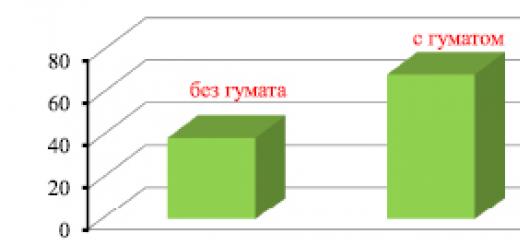
Radioactive isotopes are increasingly used in agriculture. Irradiation of plant seeds (cotton, cabbage, radishes, etc.) with small doses of gamma rays from radioactive drugs leads to a noticeable increase in yield. Large doses of radiation cause mutations in plants and microorganisms, which in some cases leads to the appearance of mutants with new valuable properties (radio selection). This is how valuable varieties of wheat, beans and other crops were developed, and highly productive microorganisms used in the production of antibiotics were obtained. Gamma radiation from radioactive isotopes is also used to combat harmful insects and for food preservation. “Tagged atoms" are widely used in agricultural technology. For example, to find out which phosphorus fertilizer is better absorbed by the plant, various fertilizers are labeled with radioactive phosphorus 15 32P. Researching Then the plants are tested for radioactivity, and the amount of phosphorus they have absorbed from different types of fertilizer can be determined.
An interesting application of radioactivity is the method of dating archaeological and geological finds by the concentration of radioactive isotopes. The most commonly used method of dating is radiocarbon dating. An unstable isotope of carbon appears in the atmosphere due to nuclear reactions caused by cosmic rays. A small percentage of this isotope is found in the air along with the usual stable isotope. Plants and other organisms take up carbon from the air, and both isotopes accumulate in them in the same proportion as in the air. After the plants die, they stop consuming carbon and the unstable isotope gradually turns into nitrogen as a result of β-decay with a half-life of 5730 years. By accurately measuring the relative concentration of radioactive carbon in the remains of ancient organisms, the time of their death can be determined.
List of used literature
1. The doctrine of radioactivity. History and modernity. M. Nauka, 1973 2. Nuclear radiation in science and technology. M. Nauka, 1984 Furman V.I. 3. Alpha decay and related nuclear reactions. M. Nauka, 1985
4. Landsberg G.S. Elementary textbook of physics. Volume III. – M.: Nauka, 19865. Seleznev Yu. A. Fundamentals of elementary physics. –M.: Nauka, 1964.6. CD ROM "Big Encyclopedia of Cyril and Methodius", 1997.
7. Curie M., Radioactivity, trans. from French, 2nd ed., M. - L., 1960
8. Murin A.N., Introduction to radioactivity, Leningrad, 1955
9. Davydov A.S., Theory of the atomic nucleus, M., 1958
10. Gaisinsky M.N., Nuclear chemistry and its applications, trans. from French, M., 1961
11. Experimental Nuclear Physics, ed. E. Segre, trans. from English, vol. 3, M., 1961; INTERNET tools